4BJH

Crystal Structure of the Aquifex Reactor Complex Formed by Dihydroorotase (H180A, H232A) with Dihydroorotate and Aspartate Transcarbamoylase with N-(phosphonacetyl)-L-aspartate (PALA)


Experimental Data Snapshot

  • Method: X-RAY DIFFRACTION
  • Resolution: 2.20 Å
  • R-Value Free: 0.203 
  • R-Value Work: 0.161 
  • R-Value Observed: 0.163 

wwPDB Validation   3D Report Full Report


Ligand Structure Quality Assessment 


This is version 1.3 of the entry. See complete history


Literature

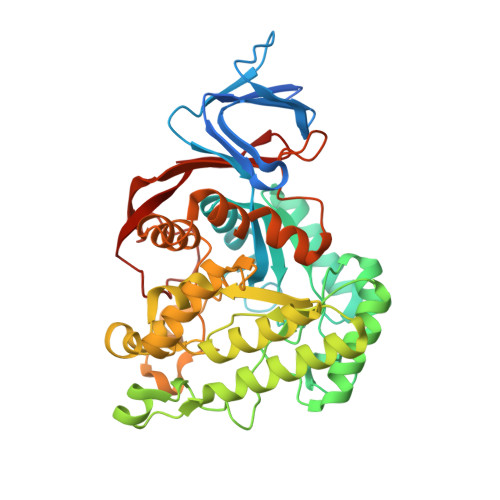
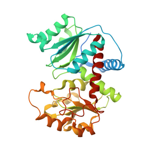
The Mononuclear Metal Center of Type-I Dihydroorotase from Aquifex Aeolicus.

Edwards, B.F.Fernando, R.Martin, P.D.Grimley, E.Cordes, M.Vaishnav, A.Brunzelle, J.S.Evans, H.G.Evans, D.R.

(2013) BMC Biochem 14: 36

  • DOI: https://doi.org/10.1186/1471-2091-14-36
  • Primary Citation of Related Structures:  
    4BJH

  • PubMed Abstract: 

    Dihydroorotase (DHO) is a zinc metalloenzyme, although the number of active site zinc ions has been controversial. E. coli DHO was initially thought to have a mononuclear metal center, but the subsequent X-ray structure clearly showed two zinc ions, α and β, at the catalytic site. Aquifex aeolicus DHO, is a dodecamer comprised of six DHO and six aspartate transcarbamoylase (ATC) subunits. The isolated DHO monomer, which lacks catalytic activity, has an intact α-site and conserved β-site ligands, but the geometry of the second metal binding site is completely disrupted. However, the putative β-site is restored when the complex with ATC is formed and DHO activity is regained. Nevertheless, the X-ray structure of the complex revealed a single zinc ion at the active site. The structure of DHO from the pathogenic organism, S. aureus showed that it also has a single active site metal ion.


  • Organizational Affiliation

    Department of Biochemistry and Molecular Biology, Wayne State University School of Medicine, 540 East Canfield Street, Detroit, MI 48201, USA. drevans@med.wayne.edu.


Macromolecules
Find similar proteins by:  (by identity cutoff)  |  3D Structure
Entity ID: 1
MoleculeChains Sequence LengthOrganismDetailsImage
DIHYDROOROTASE456Aquifex aeolicusMutation(s): 2 
EC: 3.5.2.3
UniProt
Find proteins for O66990 (Aquifex aeolicus (strain VF5))
Explore O66990 
Go to UniProtKB:  O66990
Entity Groups  
Sequence Clusters30% Identity50% Identity70% Identity90% Identity95% Identity100% Identity
UniProt GroupO66990
Sequence Annotations
Expand
  • Reference Sequence
Find similar proteins by:  (by identity cutoff)  |  3D Structure
Entity ID: 2
MoleculeChains Sequence LengthOrganismDetailsImage
ASPARTATE CARBAMOYLTRANSFERASE322Aquifex aeolicusMutation(s): 0 
EC: 2.1.3.2
UniProt
Find proteins for O66726 (Aquifex aeolicus (strain VF5))
Explore O66726 
Go to UniProtKB:  O66726
Entity Groups  
Sequence Clusters30% Identity50% Identity70% Identity90% Identity95% Identity100% Identity
UniProt GroupO66726
Sequence Annotations
Expand
  • Reference Sequence
Small Molecules
Ligands 6 Unique
IDChains Name / Formula / InChI Key2D Diagram3D Interactions
PAL
Query on PAL

Download Ideal Coordinates CCD File 
E [auth B]N-(PHOSPHONACETYL)-L-ASPARTIC ACID
C6 H10 N O8 P
ZZKNRXZVGOYGJT-VKHMYHEASA-N
DOR
Query on DOR

Download Ideal Coordinates CCD File 
D [auth A](4S)-2,6-DIOXOHEXAHYDROPYRIMIDINE-4-CARBOXYLIC ACID
C5 H6 N2 O4
UFIVEPVSAGBUSI-REOHCLBHSA-N
BA
Query on BA

Download Ideal Coordinates CCD File 
G [auth B],
H [auth B],
I [auth B]
BARIUM ION
Ba
XDFCIPNJCBUZJN-UHFFFAOYSA-N
PO4
Query on PO4

Download Ideal Coordinates CCD File 
F [auth B]PHOSPHATE ION
O4 P
NBIIXXVUZAFLBC-UHFFFAOYSA-K
ZN
Query on ZN

Download Ideal Coordinates CCD File 
C [auth A]ZINC ION
Zn
PTFCDOFLOPIGGS-UHFFFAOYSA-N
EDO
Query on EDO

Download Ideal Coordinates CCD File 
J [auth B]
K [auth B]
L [auth B]
M [auth B]
N [auth B]
J [auth B],
K [auth B],
L [auth B],
M [auth B],
N [auth B],
O [auth B],
P [auth B],
Q [auth B],
R [auth B],
S [auth B],
T [auth B],
U [auth B]
1,2-ETHANEDIOL
C2 H6 O2
LYCAIKOWRPUZTN-UHFFFAOYSA-N
Experimental Data & Validation

Experimental Data

  • Method: X-RAY DIFFRACTION
  • Resolution: 2.20 Å
  • R-Value Free: 0.203 
  • R-Value Work: 0.161 
  • R-Value Observed: 0.163 
  • Space Group: H 3 2
Unit Cell:
Length ( Å )Angle ( ˚ )
a = 157.152α = 90
b = 157.152β = 90
c = 233.244γ = 120
Software Package:
Software NamePurpose
REFMACrefinement
MOSFLMdata reduction
SCALEPACKdata scaling
ARP/wARPphasing

Structure Validation

View Full Validation Report


Ligand Structure Quality Assessment 


Entry History 

Deposition Data

Revision History  (Full details and data files)

  • Version 1.0: 2013-12-18
    Type: Initial release
  • Version 1.1: 2017-06-28
    Changes: Refinement description
  • Version 1.2: 2019-05-29
    Changes: Data collection, Experimental preparation
  • Version 1.3: 2023-12-20
    Changes: Data collection, Database references, Derived calculations, Other, Refinement description