4BJ9

Crystal structure of human tankyrase 2 in complex with EB-47


Experimental Data Snapshot

  • Method: X-RAY DIFFRACTION
  • Resolution: 2.05 Å
  • R-Value Free: 0.197 
  • R-Value Work: 0.171 
  • R-Value Observed: 0.173 

wwPDB Validation   3D Report Full Report


Ligand Structure Quality Assessment 


This is version 1.2 of the entry. See complete history


Literature

Evaluation and Structural Basis for the Inhibition of Tankyrases by Parp Inhibitors

Haikarainen, T.Narwal, M.Joensuu, P.Lehtio, L.

(2014) ACS Med Chem Lett 5: 18

  • DOI: https://doi.org/10.1021/ml400292s
  • Primary Citation of Related Structures:  
    4AVU, 4AVW, 4BJ9, 4BJB, 4BJC

  • PubMed Abstract: 

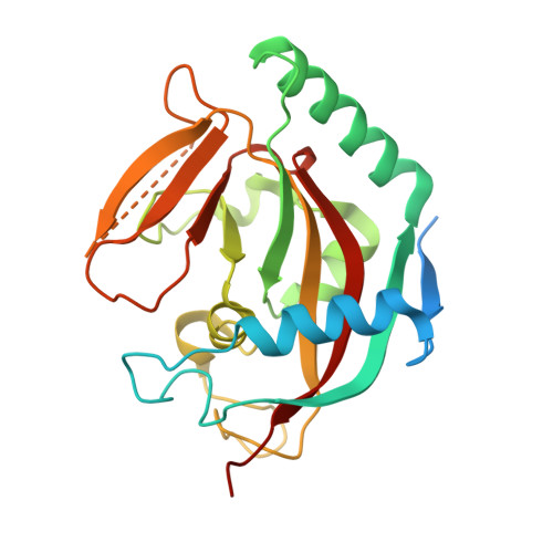
    Tankyrases, an enzyme subfamily of human poly(ADP-ribosyl)polymerases, are potential drug targets especially against cancer. We have evaluated inhibition of tankyrases by known PARP inhibitors and report five cocrystal structures of the most potent compounds in complex with human tankyrase 2. The inhibitors include the small general PARP inhibitors Phenanthridinone, PJ-34, and TIQ-A as well as the more advanced inhibitors EB-47 and rucaparib. The compounds anchor to the nicotinamide subsite of tankyrase 2. Crystal structures reveal flexibility of the ligand binding site with implications for drug development against tankyrases and other ADP-ribosyltransferases. EB-47 mimics the substrate NAD(+) and extends from the nicotinamide to the adenosine subsite. The clinical ARTD1 inhibitor candidate rucaparib was the most potent tankyrase inhibitor identified (24 and 14 nM for tankyrases), which indicates that inhibition of tankyrases would affect the cellular responses of this compound.


  • Organizational Affiliation

    Biocenter Oulu, Department of Biochemistry, University of Oulu , Oulu, Finland.


Macromolecules
Find similar proteins by:  (by identity cutoff)  |  3D Structure
Entity ID: 1
MoleculeChains Sequence LengthOrganismDetailsImage
TANKYRASE-2
A, B
240Homo sapiensMutation(s): 0 
EC: 2.4.2.30
UniProt & NIH Common Fund Data Resources
Find proteins for Q9H2K2 (Homo sapiens)
Explore Q9H2K2 
Go to UniProtKB:  Q9H2K2
PHAROS:  Q9H2K2
GTEx:  ENSG00000107854 
Entity Groups  
Sequence Clusters30% Identity50% Identity70% Identity90% Identity95% Identity100% Identity
UniProt GroupQ9H2K2
Sequence Annotations
Expand
  • Reference Sequence
Small Molecules
Ligands 4 Unique
IDChains Name / Formula / InChI Key2D Diagram3D Interactions
UHB
Query on UHB

Download Ideal Coordinates CCD File 
F [auth A],
K [auth B]
2-[4-[(2S,3S,4R,5R)-5-(6-aminopurin-9-yl)-3,4-bis(oxidanyl)oxolan-2-yl]carbonylpiperazin-1-yl]-N-(1-oxidanylidene-2,3-dihydroisoindol-4-yl)ethanamide
C24 H27 N9 O6
QEUOCRGVJJDDTK-VBFAUCSFSA-N
SO4
Query on SO4

Download Ideal Coordinates CCD File 
D [auth A],
E [auth A],
I [auth B],
J [auth B]
SULFATE ION
O4 S
QAOWNCQODCNURD-UHFFFAOYSA-L
GOL
Query on GOL

Download Ideal Coordinates CCD File 
G [auth A],
L [auth B]
GLYCEROL
C3 H8 O3
PEDCQBHIVMGVHV-UHFFFAOYSA-N
ZN
Query on ZN

Download Ideal Coordinates CCD File 
C [auth A],
H [auth B]
ZINC ION
Zn
PTFCDOFLOPIGGS-UHFFFAOYSA-N
Binding Affinity Annotations 
IDSourceBinding Affinity
UHB PDBBind:  4BJ9 IC50: 45 (nM) from 1 assay(s)
Binding MOAD:  4BJ9 IC50: 45 (nM) from 1 assay(s)
Experimental Data & Validation

Experimental Data

  • Method: X-RAY DIFFRACTION
  • Resolution: 2.05 Å
  • R-Value Free: 0.197 
  • R-Value Work: 0.171 
  • R-Value Observed: 0.173 
  • Space Group: C 2 2 21
Unit Cell:
Length ( Å )Angle ( ˚ )
a = 90.56α = 90
b = 97.78β = 90
c = 120.03γ = 90
Software Package:
Software NamePurpose
REFMACrefinement
XDSdata reduction
XSCALEdata scaling

Structure Validation

View Full Validation Report


Ligand Structure Quality Assessment 


Entry History 

Deposition Data

Revision History  (Full details and data files)

  • Version 1.0: 2013-12-11
    Type: Initial release
  • Version 1.1: 2014-06-11
    Changes: Database references
  • Version 1.2: 2014-06-18
    Changes: Database references