4BGF

The 3D-structure of arylamine-N-acetyltransferase from M. tuberculosis


Experimental Data Snapshot

  • Method: X-RAY DIFFRACTION
  • Resolution: 2.10 Å
  • R-Value Free: 0.187 
  • R-Value Work: 0.161 
  • R-Value Observed: 0.163 

wwPDB Validation   3D Report Full Report


This is version 2.0 of the entry. See complete history


Literature

Structure of arylamine N-acetyltransferase from Mycobacterium tuberculosis determined by cross-seeding with the homologous protein from M. marinum: triumph over adversity.

Abuhammad, A.Lowe, E.D.McDonough, M.A.Shaw Stewart, P.D.Kolek, S.A.Sim, E.Garman, E.F.

(2013) Acta Crystallogr D Biol Crystallogr 69: 1433-1446

  • DOI: https://doi.org/10.1107/S0907444913015126
  • Primary Citation of Related Structures:  
    4BGF

  • PubMed Abstract: 

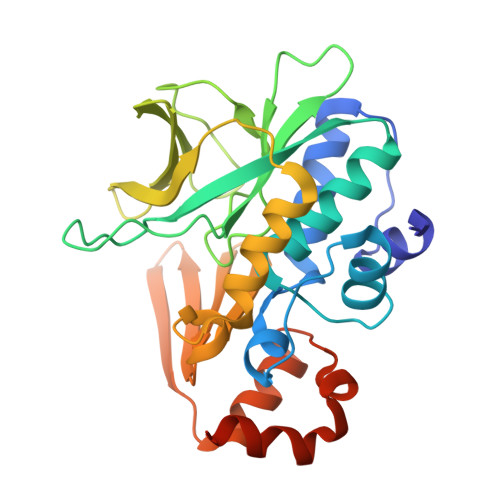
    Arylamine N-acetyltransferase from Mycobacterium tuberculosis (TBNAT) plays an important role in the intracellular survival of the microorganism inside macrophages. Medicinal chemistry efforts to optimize inhibitors of the TBNAT enzyme have been hampered by the lack of a three-dimensional structure of the enzyme. In this paper, the first structure of TBNAT, determined using a lone crystal produced using cross-seeding with the homologous protein from M. marinum, is reported. Despite the similarity between the two enzymes (74% sequence identity), they show distinct physical and biochemical characteristics. The structure elegantly reveals the characteristic features of the protein surface as well as details of the active site of TBNAT relevant to drug-discovery efforts. The crystallographic analysis of the diffraction data presented many challenges, since the crystal was twinned and the habit possessed pseudo-translational symmetry.


  • Organizational Affiliation

    Department of Pharmacology, University of Oxford, Mansfield Road, Oxford OX1 3QT, England.


Macromolecules
Find similar proteins by:  (by identity cutoff)  |  3D Structure
Entity ID: 1
MoleculeChains Sequence LengthOrganismDetailsImage
ARYLAMINE N-ACETYLTRANSFERASE NAT
A, B, C, D, E
A, B, C, D, E, F, G, H
283Mycobacterium tuberculosis H37RvMutation(s): 0 
EC: 2.3.1.5
Entity Groups  
Sequence Clusters30% Identity50% Identity70% Identity90% Identity95% Identity100% Identity
Sequence Annotations
Expand
  • Reference Sequence
Experimental Data & Validation

Experimental Data

  • Method: X-RAY DIFFRACTION
  • Resolution: 2.10 Å
  • R-Value Free: 0.187 
  • R-Value Work: 0.161 
  • R-Value Observed: 0.163 
  • Space Group: P 1 21 1
Unit Cell:
Length ( Å )Angle ( ˚ )
a = 96.494α = 90
b = 139.147β = 91.18
c = 96.506γ = 90
Software Package:
Software NamePurpose
PHENIXrefinement
xia2data reduction
XDSdata scaling
PHASERphasing

Structure Validation

View Full Validation Report


Entry History 

Deposition Data

Revision History  (Full details and data files)

  • Version 1.0: 2013-07-24
    Type: Initial release
  • Version 1.1: 2013-07-31
    Changes: Atomic model, Database references
  • Version 1.2: 2013-08-07
    Changes: Database references
  • Version 1.3: 2013-11-13
    Changes: Atomic model
  • Version 1.4: 2018-03-14
    Changes: Database references
  • Version 2.0: 2023-12-20
    Changes: Atomic model, Data collection, Database references, Other, Refinement description