4B93

Complex of Vamp7 cytoplasmic domain with 2nd ankyrin repeat domain of Varp


Experimental Data Snapshot

  • Method: X-RAY DIFFRACTION
  • Resolution: 2.00 Å
  • R-Value Free: 0.251 
  • R-Value Work: 0.213 
  • R-Value Observed: 0.215 

wwPDB Validation   3D Report Full Report


This is version 1.4 of the entry. See complete history


Literature

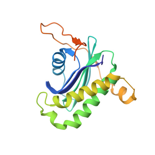
The Binding of Varp to Vamp7 Traps Vamp7 in a Closed, Fusogenically Inactive Conformation.

Schafer, I.B.Hesketh, G.G.Bright, N.A.Gray, S.R.Pryor, P.R.Evans, P.R.Luzio, J.P.Owen, D.J.

(2012) Nat Struct Mol Biol 19: 1300

  • DOI: https://doi.org/10.1038/nsmb.2414
  • Primary Citation of Related Structures:  
    4B93

  • PubMed Abstract: 

    SNAREs provide energy and specificity to membrane fusion events. Fusogenic trans-SNARE complexes are assembled from glutamine-contributing SNAREs (Q-SNAREs) embedded in one membrane and an arginine-contributing SNARE (R-SNARE) embedded in the other. Regulation of membrane fusion events is crucial for intracellular trafficking. We identify the endosomal protein Varp as an R-SNARE-binding regulator of SNARE complex formation. Varp colocalizes with and binds to VAMP7, an R-SNARE that is involved in both endocytic and secretory pathways. We present the structure of the second ankyrin repeat domain of mammalian Varp in complex with the cytosolic portion of VAMP7. The VAMP7-SNARE motif is trapped between Varp and the VAMP7 longin domain, and hence Varp kinetically inhibits the ability of VAMP7 to form SNARE complexes. This inhibition will be increased when Varp can also bind to other proteins present on the same membrane as VAMP7, such as Rab32-GTP.


  • Organizational Affiliation

    Medical Research Council Laboratory of Molecular Biology, Cambridge, UK.


Macromolecules
Find similar proteins by:  (by identity cutoff)  |  3D Structure
Entity ID: 1
MoleculeChains Sequence LengthOrganismDetailsImage
VESICLE-ASSOCIATED MEMBRANE PROTEIN 7189Mus musculusMutation(s): 0 
UniProt
Find proteins for P70280 (Mus musculus)
Explore P70280 
Go to UniProtKB:  P70280
Entity Groups  
Sequence Clusters30% Identity50% Identity70% Identity90% Identity95% Identity100% Identity
UniProt GroupP70280
Sequence Annotations
Expand
  • Reference Sequence
Find similar proteins by:  (by identity cutoff)  |  3D Structure
Entity ID: 2
MoleculeChains Sequence LengthOrganismDetailsImage
ANKYRIN REPEAT DOMAIN-CONTAINING PROTEIN 27269Homo sapiensMutation(s): 0 
UniProt & NIH Common Fund Data Resources
Find proteins for Q96NW4 (Homo sapiens)
Explore Q96NW4 
Go to UniProtKB:  Q96NW4
PHAROS:  Q96NW4
GTEx:  ENSG00000105186 
Entity Groups  
Sequence Clusters30% Identity50% Identity70% Identity90% Identity95% Identity100% Identity
UniProt GroupQ96NW4
Sequence Annotations
Expand
  • Reference Sequence
Experimental Data & Validation

Experimental Data

  • Method: X-RAY DIFFRACTION
  • Resolution: 2.00 Å
  • R-Value Free: 0.251 
  • R-Value Work: 0.213 
  • R-Value Observed: 0.215 
  • Space Group: C 2 2 21
Unit Cell:
Length ( Å )Angle ( ˚ )
a = 69.57α = 90
b = 122.66β = 90
c = 158.6γ = 90
Software Package:
Software NamePurpose
REFMACrefinement
MOSFLMdata reduction
Aimlessdata scaling
SHARPphasing

Structure Validation

View Full Validation Report


Entry History 

Deposition Data

Revision History  (Full details and data files)

  • Version 1.0: 2012-10-31
    Type: Initial release
  • Version 1.1: 2012-12-19
    Changes: Database references
  • Version 1.2: 2017-01-25
    Changes: Data collection
  • Version 1.3: 2019-04-03
    Changes: Data collection, Experimental preparation, Other
  • Version 1.4: 2019-05-08
    Changes: Data collection, Experimental preparation