4B8Y

Ferrichrome-bound FhuD2


Experimental Data Snapshot

  • Method: X-RAY DIFFRACTION
  • Resolution: 1.90 Å
  • R-Value Free: 0.223 
  • R-Value Work: 0.174 
  • R-Value Observed: 0.176 

wwPDB Validation   3D Report Full Report


This is version 2.1 of the entry. See complete history


Literature

Structural and Functional Characterization of the Staphylococcus Aureus Virulence Factor and Vaccine Candidate Fhud2.

Mariotti, P.Malito, E.Biancucci, M.Lo Surdo, P.Mishra, R.P.Nardi-Dei, V.Savino, S.Nissum, M.Spraggon, G.Grandi, G.Bagnoli, F.Bottomley, M.J.

(2013) Biochem J 449: 683

  • DOI: https://doi.org/10.1042/BJ20121426
  • Primary Citation of Related Structures:  
    4B8Y

  • PubMed Abstract: 

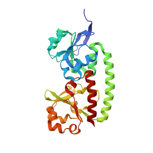
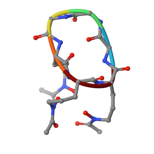
    Staphylococcus aureus is a human pathogen causing globally significant morbidity and mortality. The development of antibiotic resistance in S. aureus highlights the need for a preventive vaccine. In the present paper we explore the structure and function of FhuD2 (ferric-hydroxamate uptake D2), a staphylococcal surface lipoprotein mediating iron uptake during invasive infection, recently described as a promising vaccine candidate. Differential scanning fluorimetry and calorimetry studies revealed that FhuD2 is stabilized by hydroxamate siderophores. The FhuD2-ferrichrome interaction was of nanomolar affinity in surface plasmon resonance experiments and fully iron(III)-dependent. We determined the X-ray crystallographic structure of ligand-bound FhuD2 at 1.9 Å (1 Å=0.1 nm) resolution, revealing the bilobate fold of class III SBPs (solute-binding proteins). The ligand, ferrichrome, occupies a cleft between the FhuD2 N- and C-terminal lobes. Many FhuD2-siderophore interactions enable the specific recognition of ferrichrome. Biochemical data suggest that FhuD2 does not undergo significant conformational changes upon siderophore binding, supporting the hypothesis that the ligand-bound complex is essential for receptor engagement and uptake. Finally, immunizations with FhuD2 alone or FhuD2 formulated with hydroxamate siderophores were equally protective in a murine staphylococcal infection model, confirming the suitability and efficacy of apo-FhuD2 as a protective antigen, and suggesting that other class III SBPs might also be exploited as vaccine candidates.


  • Organizational Affiliation

    Novartis Vaccines and Diagnostics srl, Via Fiorentina 1, 53100 Siena, Italy.


Macromolecules
Find similar proteins by:  (by identity cutoff)  |  3D Structure
Entity ID: 1
MoleculeChains Sequence LengthOrganismDetailsImage
FHUD2277Staphylococcus aureus subsp. aureus NCTC 8325Mutation(s): 0 
UniProt
Find proteins for Q2FVW9 (Staphylococcus aureus (strain NCTC 8325 / PS 47))
Explore Q2FVW9 
Go to UniProtKB:  Q2FVW9
Entity Groups  
Sequence Clusters30% Identity50% Identity70% Identity90% Identity95% Identity100% Identity
UniProt GroupQ2FVW9
Sequence Annotations
Expand
  • Reference Sequence

Find similar proteins by:  Sequence   |   3D Structure  

Entity ID: 2
MoleculeChains Sequence LengthOrganismDetailsImage
VIRULENCE FACTOR6synthetic constructMutation(s): 0 
Sequence Annotations
Expand
  • Reference Sequence
Small Molecules
Ligands 3 Unique
IDChains Name / Formula / InChI Key2D Diagram3D Interactions
EDO
Query on EDO

Download Ideal Coordinates CCD File 
C [auth A]
D [auth A]
E [auth A]
F [auth A]
G [auth A]
C [auth A],
D [auth A],
E [auth A],
F [auth A],
G [auth A],
H [auth A],
I [auth A],
J [auth A],
K [auth A],
L [auth A]
1,2-ETHANEDIOL
C2 H6 O2
LYCAIKOWRPUZTN-UHFFFAOYSA-N
FE
Query on FE

Download Ideal Coordinates CCD File 
N [auth B]FE (III) ION
Fe
VTLYFUHAOXGGBS-UHFFFAOYSA-N
CL
Query on CL

Download Ideal Coordinates CCD File 
M [auth A]CHLORIDE ION
Cl
VEXZGXHMUGYJMC-UHFFFAOYSA-M
Modified Residues  1 Unique
IDChains TypeFormula2D DiagramParent
AHO
Query on AHO
B
L-PEPTIDE LINKINGC7 H14 N2 O4ALA
Experimental Data & Validation

Experimental Data

  • Method: X-RAY DIFFRACTION
  • Resolution: 1.90 Å
  • R-Value Free: 0.223 
  • R-Value Work: 0.174 
  • R-Value Observed: 0.176 
  • Space Group: P 1 21 1
Unit Cell:
Length ( Å )Angle ( ˚ )
a = 53.89α = 90
b = 50.14β = 106.44
c = 54.35γ = 90
Software Package:
Software NamePurpose
PHENIXrefinement
MOSFLMdata reduction
SCALAdata scaling
PHASERphasing

Structure Validation

View Full Validation Report


Entry History 

Deposition Data

Revision History  (Full details and data files)

  • Version 1.0: 2012-11-21
    Type: Initial release
  • Version 1.1: 2012-12-05
    Changes: Database references
  • Version 1.2: 2013-01-23
    Changes: Database references
  • Version 2.0: 2023-11-15
    Changes: Atomic model, Data collection, Database references, Derived calculations, Other
  • Version 2.1: 2023-12-20
    Changes: Refinement description