4B86

Crystal structure of the MSL1-MSL2 complex (3.5A)


Experimental Data Snapshot

  • Method: X-RAY DIFFRACTION
  • Resolution: 3.50 Å
  • R-Value Free: 0.297 
  • R-Value Work: 0.256 
  • R-Value Observed: 0.258 

wwPDB Validation   3D Report Full Report


This is version 1.0 of the entry. See complete history


Literature

Msl1-Mediated Dimerization of the Dosage Compensation Complex is Essential for Male X-Chromosome Regulation in Drosophila.

Hallacli, E.Lipp, M.Georgiev, P.Spielman, C.Cusack, S.Akhtar, A.Kadlec, J.

(2012) Mol Cell 48: 587

  • DOI: https://doi.org/10.1016/j.molcel.2012.09.014
  • Primary Citation of Related Structures:  
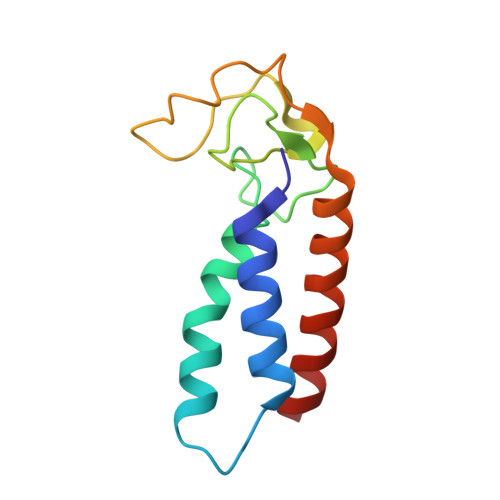
    4B7Y, 4B86

  • PubMed Abstract: 

    The Male-Specific Lethal (MSL) complex regulates dosage compensation of the male X chromosome in Drosophila. Here, we report the crystal structure of its MSL1/MSL2 core, where two MSL2 subunits bind to a dimer formed by two molecules of MSL1. Analysis of structure-based mutants revealed that MSL2 can only interact with the MSL1 dimer, but MSL1 dimerization is MSL2 independent. We show that Msl1 is a substrate for Msl2 E3 ubiquitin ligase activity. ChIP experiments revealed that Msl1 dimerization is essential for targeting and spreading of the MSL complex on X-linked genes; however, Msl1 binding to promoters of male and female cells is independent of the dimer status and other MSL proteins. Finally, we show that loss of Msl1 dimerization leads to male-specific lethality. We propose that Msl1-mediated dimerization of the entire MSL complex is required for Msl2 binding, X chromosome recognition, and spreading along the X chromosome.


  • Organizational Affiliation

    Max-Planck-Institut für Immunbiologie und Epigenetik, Stübeweg 51, 79108 Freiburg im Breisgau, Germany.


Macromolecules
Find similar proteins by:  (by identity cutoff)  |  3D Structure
Entity ID: 1
MoleculeChains Sequence LengthOrganismDetailsImage
MALE-SPECIFIC LETHAL 1 HOMOLOG
A, B, E, F, I
A, B, E, F, I, J
59Homo sapiensMutation(s): 0 
UniProt & NIH Common Fund Data Resources
Find proteins for Q68DK7 (Homo sapiens)
Explore Q68DK7 
Go to UniProtKB:  Q68DK7
PHAROS:  Q68DK7
GTEx:  ENSG00000188895 
Entity Groups  
Sequence Clusters30% Identity50% Identity70% Identity90% Identity95% Identity100% Identity
UniProt GroupQ68DK7
Sequence Annotations
Expand
  • Reference Sequence
Find similar proteins by:  (by identity cutoff)  |  3D Structure
Entity ID: 2
MoleculeChains Sequence LengthOrganismDetailsImage
MALE-SPECIFIC LETHAL 2 HOMOLOG
C, D, G, H, K
C, D, G, H, K, L
116Homo sapiensMutation(s): 0 
UniProt & NIH Common Fund Data Resources
Find proteins for Q9HCI7 (Homo sapiens)
Explore Q9HCI7 
Go to UniProtKB:  Q9HCI7
PHAROS:  Q9HCI7
GTEx:  ENSG00000174579 
Entity Groups  
Sequence Clusters30% Identity50% Identity70% Identity90% Identity95% Identity100% Identity
UniProt GroupQ9HCI7
Sequence Annotations
Expand
  • Reference Sequence
Small Molecules
Ligands 1 Unique
IDChains Name / Formula / InChI Key2D Diagram3D Interactions
ZN
Query on ZN

Download Ideal Coordinates CCD File 
M [auth C]
N [auth C]
O [auth D]
P [auth D]
Q [auth G]
M [auth C],
N [auth C],
O [auth D],
P [auth D],
Q [auth G],
R [auth G],
S [auth H],
T [auth H],
U [auth K],
V [auth K],
W [auth L],
X [auth L]
ZINC ION
Zn
PTFCDOFLOPIGGS-UHFFFAOYSA-N
Experimental Data & Validation

Experimental Data

  • Method: X-RAY DIFFRACTION
  • Resolution: 3.50 Å
  • R-Value Free: 0.297 
  • R-Value Work: 0.256 
  • R-Value Observed: 0.258 
  • Space Group: P 21 21 2
Unit Cell:
Length ( Å )Angle ( ˚ )
a = 104.65α = 90
b = 182.21β = 90
c = 89.47γ = 90
Software Package:
Software NamePurpose
XDSdata reduction
XSCALEdata scaling
SHELXDphasing
SHARPphasing
REFMACrefinement

Structure Validation

View Full Validation Report


Entry History 

Deposition Data

Revision History  (Full details and data files)

  • Version 1.0: 2013-02-06
    Type: Initial release