


4B81
Mus musculus Acetylcholinesterase in complex with 1-(4-Chloro-phenyl)- N-(2-diethylamino-ethyl)-methanesulfonamide
- PDB DOI: https://doi.org/10.2210/pdb4B81/pdb
- Classification: HYDROLASE
- Organism(s): Mus musculus
- Expression System: Homo sapiens
- Mutation(s): No
- Deposited: 2012-08-24 Released: 2013-09-04
Experimental Data Snapshot
- Method: X-RAY DIFFRACTION
- Resolution: 2.80 Å
- R-Value Free: 0.239
- R-Value Work: 0.197
- R-Value Observed: 0.197

This is version 2.2 of the entry. See complete history.

Macromolecules
Find similar proteins by:
(by identity cutoff) | 3D Structure
Entity ID: 1 | |||||
|---|---|---|---|---|---|
| Molecule | Chains | Sequence Length | Organism | Details | Image |
| ACETYLCHOLINESTERASE | 548 | Mus musculus | Mutation(s): 0 EC: 3.1.1.7 | 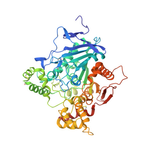 | |
UniProt & NIH Common Fund Data Resources | |||||
Find proteins for P21836 (Mus musculus) Explore P21836 Go to UniProtKB: P21836 | |||||
IMPC: MGI:87876 | |||||
Entity Groups | |||||
| Sequence Clusters | 30% Identity50% Identity70% Identity90% Identity95% Identity100% Identity | ||||
| UniProt Group | P21836 | ||||
Sequence AnnotationsExpand | |||||
| |||||
Small Molecules
| Ligands 5 Unique | |||||
|---|---|---|---|---|---|
| ID | Chains | Name / Formula / InChI Key | 2D Diagram | 3D Interactions | |
| ZN4 Query on ZN4 | C [auth A], K [auth B] | 1-(4-chlorophenyl)-N-[2-(diethylamino)ethyl]methanesulfonamide C13 H21 Cl N2 O2 S MKQJIDUAXALKCV-UHFFFAOYSA-N |  | ||
| P6G Query on P6G | J [auth B] | HEXAETHYLENE GLYCOL C12 H26 O7 IIRDTKBZINWQAW-UHFFFAOYSA-N |  | ||
| NAG Query on NAG | E [auth A], G [auth A], P [auth B] | 2-acetamido-2-deoxy-beta-D-glucopyranose C8 H15 N O6 OVRNDRQMDRJTHS-FMDGEEDCSA-N |  | ||
| PEG Query on PEG | F [auth A] H [auth A] I [auth A] L [auth B] N [auth B] | DI(HYDROXYETHYL)ETHER C4 H10 O3 MTHSVFCYNBDYFN-UHFFFAOYSA-N |  | ||
| SO4 Query on SO4 | D [auth A], M [auth B] | SULFATE ION O4 S QAOWNCQODCNURD-UHFFFAOYSA-L |  | ||
Experimental Data & Validation
Experimental Data
- Method: X-RAY DIFFRACTION
- Resolution: 2.80 Å
- R-Value Free: 0.239
- R-Value Work: 0.197
- R-Value Observed: 0.197
- Space Group: P 21 21 21
Unit Cell:
| Length ( Å ) | Angle ( ˚ ) |
|---|---|
| a = 77.959 | α = 90 |
| b = 109.876 | β = 90 |
| c = 227.926 | γ = 90 |
| Software Name | Purpose |
|---|---|
| PHENIX | refinement |
| XDS | data reduction |
| SCALA | data scaling |
| REFMAC | phasing |
Entry History
Deposition Data
- Released Date: 2013-09-04 Deposition Author(s): Andersson, C.D., Forsgren, N., Akfur, C., Allgardsson, A., Berg, L., Qian, W., Ekstrom, F., Linusson, A.
Revision History (Full details and data files)
- Version 1.0: 2013-09-04
Type: Initial release - Version 1.1: 2013-09-11
Changes: Database references - Version 1.2: 2013-10-30
Changes: Database references - Version 2.0: 2018-01-17
Changes: Atomic model, Data collection - Version 2.1: 2020-07-29
Type: Remediation
Reason: Carbohydrate remediation
Changes: Advisory, Data collection, Derived calculations, Other, Structure summary - Version 2.2: 2023-12-20
Changes: Data collection, Database references, Refinement description, Structure summary
