Mechanistic basis of the inhibition of type II dehydroquinase by (2S)- and (2R)-2-benzyl-3-dehydroquinic acids.
Lence, E., Tizon, L., Otero, J.M., Peon, A., Prazeres, V.F., Llamas-Saiz, A.L., Fox, G.C., van Raaij, M.J., Lamb, H., Hawkins, A.R., Gonzalez-Bello, C.(2013) ACS Chem Biol 8: 568-577
- PubMed: 23198883
- DOI: https://doi.org/10.1021/cb300493s
- Primary Citation of Related Structures:
4B6O, 4B6P, 4B6Q, 4B6R, 4B6S - PubMed Abstract:
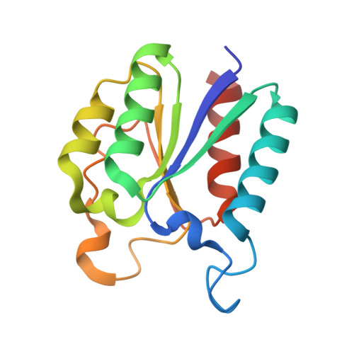
The structural changes caused by the substitution of the aromatic moiety in (2S)-2-benzyl-3-dehydroquinic acids and its epimers in C2 by electron-withdrawing or electron-donating groups in type II dehydroquinase enzyme from M. tuberculosis and H. pylori has been investigated by structural and computational studies. Both compounds are reversible competitive inhibitors of this enzyme, which is essential in these pathogenic bacteria. The crystal structures of M. tuberculosis and H. pylori in complex with (2S)-2-(4-methoxy)benzyl- and (2S)-2-perfluorobenzyl-3-dehydroquinic acids have been solved at 2.0, 2.3, 2.0, and 1.9 Å, respectively. The crystal structure of M. tuberculosis in complex with (2R)-2-(benzothiophen-5-yl)methyl-3-dehydroquinic acid is also reported at 1.55 Å. These crystal structures reveal key differences in the conformation of the flexible loop of the two enzymes, a difference that depends on the presence of electron-withdrawing or electron-donating groups in the aromatic moiety of the inhibitors. This loop closes over the active site after substrate binding, and its flexibility is essential for the function of the enzyme. These differences have also been investigated by molecular dynamics simulations in an effort to understand the significant inhibition potency differences observed between some of these compounds and also to obtain more information about the possible movements of the loop. These computational studies have also allowed us to identify key structural factors of the H. pylori loop that could explain its reduced flexibility in comparison to the M. tuberculosis loop, specifically by the formation of a key salt bridge between the side chains of residues Asp18 and Arg20.
Organizational Affiliation:
Centro Singular de Investigación en Química Biológica y Materiales Moleculares (CIQUS), Edificio CACTUS, Universidad de Santiago de Compostela, 15782 Santiago de Compostela, Spain.