4B0Z

Crystal structure of S. pombe Rpn12


Experimental Data Snapshot

  • Method: X-RAY DIFFRACTION
  • Resolution: 1.58 Å
  • R-Value Free: 0.225 
  • R-Value Work: 0.188 
  • R-Value Observed: 0.190 

wwPDB Validation   3D Report Full Report


This is version 1.1 of the entry. See complete history


Literature

Structural and Functional Characterisation of Rpn12 Identifies Residues Required for Rpn10 Proteasome Incorporation.

Boehringer, J.Riedinger, C.Paraskevopoulos, K.Johnson, E.O.D.Lowe, E.D.Khoudian, C.Smith, D.Noble, M.E.M.Gordon, C.Endicott, J.A.

(2012) Biochem J 448: 55

  • DOI: https://doi.org/10.1042/BJ20120542
  • Primary Citation of Related Structures:  
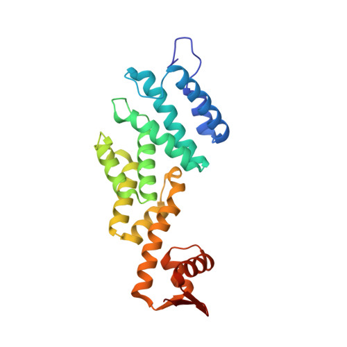
    4B0Z

  • PubMed Abstract: 

    The ubiquitin-proteasome system targets selected proteins for degradation by the 26S proteasome. Rpn12 is an essential component of the 19S regulatory particle and plays a role in recruiting the extrinsic ubiquitin receptor Rpn10. In the present paper we report the crystal structure of Rpn12, a proteasomal PCI-domain-containing protein. The structure helps to define a core structural motif for the PCI domain and identifies potential sites through which Rpn12 might form protein-protein interactions. We demonstrate that mutating residues at one of these sites impairs Rpn12 binding to Rpn10 in vitro and reduces Rpn10 incorporation into proteasomes in vivo.


  • Organizational Affiliation

    Department of Biochemistry, University of Oxford, Oxford OX1 3QU, UK.


Macromolecules
Find similar proteins by:  (by identity cutoff)  |  3D Structure
Entity ID: 1
MoleculeChains Sequence LengthOrganismDetailsImage
26S PROTEASOME REGULATORY SUBUNIT RPN12
A, B
229Schizosaccharomyces pombeMutation(s): 0 
UniProt
Find proteins for P50524 (Schizosaccharomyces pombe (strain 972 / ATCC 24843))
Explore P50524 
Go to UniProtKB:  P50524
Entity Groups  
Sequence Clusters30% Identity50% Identity70% Identity90% Identity95% Identity100% Identity
UniProt GroupP50524
Sequence Annotations
Expand
  • Reference Sequence
Small Molecules
Ligands 3 Unique
IDChains Name / Formula / InChI Key2D Diagram3D Interactions
SGM
Query on SGM

Download Ideal Coordinates CCD File 
F [auth A],
H [auth B]
MONOTHIOGLYCEROL
C3 H8 O2 S
PJUIMOJAAPLTRJ-GSVOUGTGSA-N
GOL
Query on GOL

Download Ideal Coordinates CCD File 
I [auth B],
J [auth B],
K [auth B]
GLYCEROL
C3 H8 O3
PEDCQBHIVMGVHV-UHFFFAOYSA-N
NO3
Query on NO3

Download Ideal Coordinates CCD File 
C [auth A],
D [auth A],
E [auth A],
G [auth B]
NITRATE ION
N O3
NHNBFGGVMKEFGY-UHFFFAOYSA-N
Experimental Data & Validation

Experimental Data

  • Method: X-RAY DIFFRACTION
  • Resolution: 1.58 Å
  • R-Value Free: 0.225 
  • R-Value Work: 0.188 
  • R-Value Observed: 0.190 
  • Space Group: P 21 21 21
Unit Cell:
Length ( Å )Angle ( ˚ )
a = 41.78α = 90
b = 91.38β = 90
c = 143.34γ = 90
Software Package:
Software NamePurpose
PHENIXrefinement
MOSFLMdata reduction
SCALAdata scaling
SHELXphasing

Structure Validation

View Full Validation Report


Entry History 

Revision History  (Full details and data files)

  • Version 1.0: 2012-09-12
    Type: Initial release
  • Version 1.1: 2012-11-07
    Changes: Database references