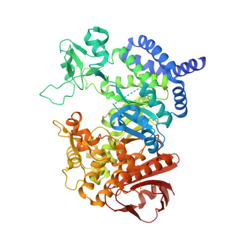
The Structure of Amylosucrase from Deinococcus Radiodurans Has an Unusual Open Active-Site Topology
Skov, L.K., Pizzut-Serin, S., Remaud-Simeon, M., Ernst, H.A., Gajhede, M., Mirza, O.(2013) Acta Crystallogr Sect F Struct Biol Cryst Commun 69: 973
- PubMed: 23989143
- DOI: https://doi.org/10.1107/S1744309113021714
- Primary Citation of Related Structures:
4AYS - PubMed Abstract:
Amylosucrases (ASes) catalyze the formation of an α-1,4-glucosidic linkage by transferring a glucosyl unit from sucrose onto an acceptor α-1,4-glucan. To date, several ligand-bound crystal structures of wild-type and mutant ASes from Neisseria polysaccharea and Deinococcus geothermalis have been solved. These structures all display a very similar overall conformation with a deep pocket leading to the site for transglucosylation, subsite -1. This has led to speculation on how sucrose enters the active site during glucan elongation. In contrast to previous studies, the AS structure from D. radiodurans presented here has a completely empty -1 subsite. This structure is strikingly different from other AS structures, as an active-site-lining loop comprising residues Leu214-Asn225 is found in a previously unobserved conformation. In addition, a large loop harbouring the conserved active-site residues Asp133 and Tyr136 is disordered. The result of the changed loop conformations is that the active-site topology is radically changed, leaving subsite -1 exposed and partially dismantled. This structure provides novel insights into the dynamics of ASes and comprises the first structural support for an elongation mechanism that involves considerable conformational changes to modulate accessibility to the sucrose-binding site and thereby allows successive cycles of glucosyl-moiety transfer to a growing glucan chain.
Organizational Affiliation:
Novozymes A/S, Krogshøjvej 36, DK-2880 Bagsværd, Denmark.