4AU7

The structure of the Suv4-20h2 ternary complex with histone H4


Experimental Data Snapshot

  • Method: X-RAY DIFFRACTION
  • Resolution: 2.07 Å
  • R-Value Free: 0.240 
  • R-Value Work: 0.193 
  • R-Value Observed: 0.195 

wwPDB Validation   3D Report Full Report


Ligand Structure Quality Assessment 


This is version 1.2 of the entry. See complete history


Literature

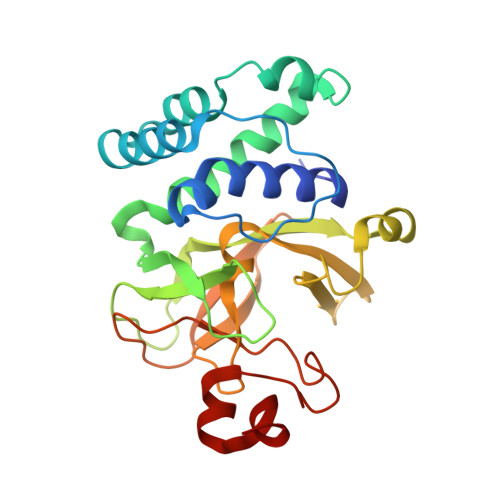
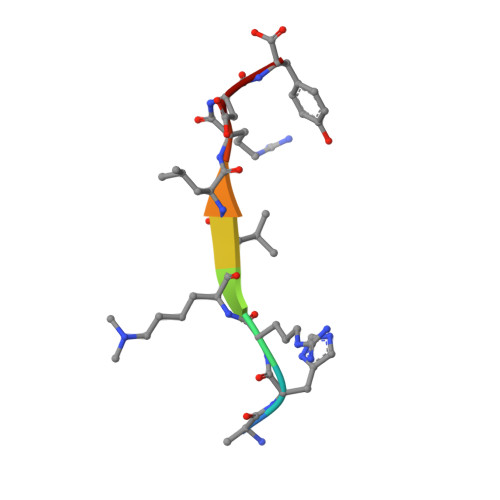
A Novel Route to Product Specificity in the Suv4-20 Family of Histone H4K20 Methyltransferases.

Southall, S.M.Cronin, N.B.Wilson, J.R.

(2014) Nucleic Acids Res 42: 661

  • DOI: https://doi.org/10.1093/nar/gkt776
  • Primary Citation of Related Structures:  
    4AU7, 4BUP

  • PubMed Abstract: 

    The delivery of site-specific post-translational modifications to histones generates an epigenetic regulatory network that directs fundamental DNA-mediated processes and governs key stages in development. Methylation of histone H4 lysine-20 has been implicated in DNA repair, transcriptional silencing, genomic stability and regulation of replication. We present the structure of the histone H4K20 methyltransferase Suv4-20h2 in complex with its histone H4 peptide substrate and S-adenosyl methionine cofactor. Analysis of the structure reveals that the Suv4-20h2 active site diverges from the canonical SET domain configuration and generates a high degree of both substrate and product specificity. Together with supporting biochemical data comparing Suv4-20h1 and Suv4-20h2, we demonstrate that the Suv4-20 family enzymes take a previously mono-methylated H4K20 substrate and generate an exclusively di-methylated product. We therefore predict that other enzymes are responsible for the tri-methylation of histone H4K20 that marks silenced heterochromatin.


  • Organizational Affiliation

    Division of Structural Biology, Institute of Cancer Research, Chester Beatty Laboratories, London, SW3 6JB, UK.


Macromolecules
Find similar proteins by:  (by identity cutoff)  |  3D Structure
Entity ID: 1
MoleculeChains Sequence LengthOrganismDetailsImage
HISTONE-LYSINE N-METHYLTRANSFERASE SUV420H2
A, B
247Mus musculusMutation(s): 0 
EC: 2.1.1.43
UniProt
Find proteins for Q6Q783 (Mus musculus)
Explore Q6Q783 
Go to UniProtKB:  Q6Q783
Entity Groups  
Sequence Clusters30% Identity50% Identity70% Identity90% Identity95% Identity100% Identity
UniProt GroupQ6Q783
Sequence Annotations
Expand
  • Reference Sequence

Find similar proteins by:  Sequence   |   3D Structure  

Entity ID: 2
MoleculeChains Sequence LengthOrganismDetailsImage
HISTONE H4 PEPTIDE9Mus musculusMutation(s): 0 
UniProt & NIH Common Fund Data Resources
Find proteins for P62806 (Mus musculus)
Explore P62806 
Go to UniProtKB:  P62806
IMPC:  MGI:2448443
Entity Groups  
UniProt GroupP62806
Sequence Annotations
Expand
  • Reference Sequence
Small Molecules
Ligands 3 Unique
IDChains Name / Formula / InChI Key2D Diagram3D Interactions
SAH
Query on SAH

Download Ideal Coordinates CCD File 
D [auth A]S-ADENOSYL-L-HOMOCYSTEINE
C14 H20 N6 O5 S
ZJUKTBDSGOFHSH-WFMPWKQPSA-N
ZN
Query on ZN

Download Ideal Coordinates CCD File 
E [auth A],
F [auth A],
O [auth B]
ZINC ION
Zn
PTFCDOFLOPIGGS-UHFFFAOYSA-N
EDO
Query on EDO

Download Ideal Coordinates CCD File 
G [auth A]
H [auth A]
I [auth A]
J [auth A]
K [auth A]
G [auth A],
H [auth A],
I [auth A],
J [auth A],
K [auth A],
L [auth A],
M [auth A],
N [auth A],
P [auth B],
Q [auth B],
R [auth B],
S [auth B],
T [auth B],
U [auth B],
V [auth B],
W [auth B]
1,2-ETHANEDIOL
C2 H6 O2
LYCAIKOWRPUZTN-UHFFFAOYSA-N
Modified Residues  1 Unique
IDChains TypeFormula2D DiagramParent
MLY
Query on MLY
C
L-PEPTIDE LINKINGC8 H18 N2 O2LYS
Binding Affinity Annotations 
IDSourceBinding Affinity
SAH PDBBind:  4AU7 Kd: 1.72e+4 (nM) from 1 assay(s)
BindingDB:  4AU7 IC50: 1.00e+4 (nM) from 1 assay(s)
Experimental Data & Validation

Experimental Data

  • Method: X-RAY DIFFRACTION
  • Resolution: 2.07 Å
  • R-Value Free: 0.240 
  • R-Value Work: 0.193 
  • R-Value Observed: 0.195 
  • Space Group: P 21 21 21
Unit Cell:
Length ( Å )Angle ( ˚ )
a = 37.27α = 90
b = 65.17β = 90
c = 209.49γ = 90
Software Package:
Software NamePurpose
REFMACrefinement
XDSdata reduction
SCALEPACKdata scaling
PHASERphasing

Structure Validation

View Full Validation Report


Ligand Structure Quality Assessment 


Entry History 

Deposition Data

Revision History  (Full details and data files)

  • Version 1.0: 2013-05-22
    Type: Initial release
  • Version 1.1: 2014-01-15
    Changes: Database references
  • Version 1.2: 2023-12-20
    Changes: Data collection, Database references, Derived calculations, Other, Refinement description