4ARK

CRYSTAL STRUCTURE OF THE CATALYTIC DOMAIN OF HUMAN MAP KINASE KINASE 1 (MEK1) IN COMPLEX WITH A SMALL MOLECULE INHIBITOR AND ADP


Experimental Data Snapshot

  • Method: X-RAY DIFFRACTION
  • Resolution: 2.60 Å
  • R-Value Free: 0.211 
  • R-Value Work: 0.172 
  • R-Value Observed: 0.174 

wwPDB Validation   3D Report Full Report


Ligand Structure Quality Assessment 


This is version 1.4 of the entry. See complete history


Literature

Optimization of Allosteric Mek Inhibitors - Part 1: Venturing Into Unexplored Sar Territories

Hartung, I.V.Hitchcock, M.Puehler, F.Neuhaus, R.Scholz, A.Hammer, S.Petersen, K.Siemeister, G.Brittain, D.Hillig, R.C.

(2013) Bioorg Med Chem Lett 23: 2384

  • DOI: https://doi.org/10.1016/j.bmcl.2013.02.028
  • Primary Citation of Related Structures:  
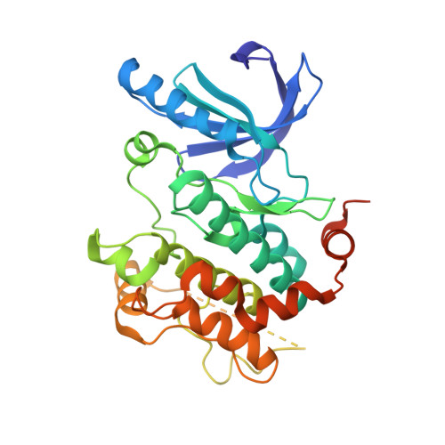
    4ARK

  • PubMed Abstract: 

    Using PD325901 as a starting point for identifying novel allosteric MEK inhibitors with high cell potency and long-lasting target inhibition in vivo, truncation of its hydroxamic ester headgroup was combined with incorporation of alkyl and aryl ethers at the neighboring ring position. Whereas alkoxy side chains did not yield sufficient levels of cell potency, specifically substituted aryloxy groups allowed for high enzymatic and cellular potencies. Sulfamide 28 was identified as a highly potent MEK inhibitor with nanomolar cell potency against B-RAF (V600E) as well as Ras-mutated cell lines, high metabolic stability and resulting long half-lives. It was efficacious against B-RAF as well as K-Ras driven xenograft models and showed-despite being orally bioavailable and not a P-glycoprotein substrate-much lower brain/plasma exposure ratios than PD325901.


  • Organizational Affiliation

    Medicinal Chemistry, Bayer HealthCare AG, 13353 Berlin, Germany. ingo.hartung@bayer.com


Macromolecules
Find similar proteins by:  (by identity cutoff)  |  3D Structure
Entity ID: 1
MoleculeChains Sequence LengthOrganismDetailsImage
DUAL SPECIFICITY MITOGEN-ACTIVATED PROTEIN KINASE KINASE 1341Homo sapiensMutation(s): 0 
EC: 2.7.12.2
UniProt & NIH Common Fund Data Resources
Find proteins for Q02750 (Homo sapiens)
Explore Q02750 
Go to UniProtKB:  Q02750
PHAROS:  Q02750
GTEx:  ENSG00000169032 
Entity Groups  
Sequence Clusters30% Identity50% Identity70% Identity90% Identity95% Identity100% Identity
UniProt GroupQ02750
Sequence Annotations
Expand
  • Reference Sequence
Small Molecules
Ligands 3 Unique
IDChains Name / Formula / InChI Key2D Diagram3D Interactions
M3K
Query on M3K

Download Ideal Coordinates CCD File 
D [auth A]2-([3R-3,4-dihydroxybutyl]oxy)-4-fluoro-6-[(2-fluoro-4-iodophenyl)amino]benzamide
C17 H17 F2 I N2 O4
HNTGGMMWHHPHAE-LLVKDONJSA-N
ADP
Query on ADP

Download Ideal Coordinates CCD File 
B [auth A]ADENOSINE-5'-DIPHOSPHATE
C10 H15 N5 O10 P2
XTWYTFMLZFPYCI-KQYNXXCUSA-N
MG
Query on MG

Download Ideal Coordinates CCD File 
C [auth A]MAGNESIUM ION
Mg
JLVVSXFLKOJNIY-UHFFFAOYSA-N
Binding Affinity Annotations 
IDSourceBinding Affinity
M3K PDBBind:  4ARK IC50: 63 (nM) from 1 assay(s)
Experimental Data & Validation

Experimental Data

  • Method: X-RAY DIFFRACTION
  • Resolution: 2.60 Å
  • R-Value Free: 0.211 
  • R-Value Work: 0.172 
  • R-Value Observed: 0.174 
  • Space Group: P 62
Unit Cell:
Length ( Å )Angle ( ˚ )
a = 82.411α = 90
b = 82.411β = 90
c = 130.238γ = 120
Software Package:
Software NamePurpose
REFMACrefinement
MOSFLMdata reduction
SCALAdata scaling
REFMACphasing

Structure Validation

View Full Validation Report


Ligand Structure Quality Assessment 


Entry History 

Deposition Data

Revision History  (Full details and data files)

  • Version 1.0: 2013-03-06
    Type: Initial release
  • Version 1.1: 2013-04-03
    Changes: Database references
  • Version 1.2: 2013-04-17
    Changes: Database references
  • Version 1.3: 2016-12-14
    Changes: Data collection
  • Version 1.4: 2023-12-20
    Changes: Data collection, Database references, Derived calculations, Other, Refinement description