4ANE

R80N MUTANT OF NUCLEOSIDE DIPHOSPHATE KINASE FROM MYCOBACTERIUM TUBERCULOSIS


Experimental Data Snapshot

  • Method: X-RAY DIFFRACTION
  • Resolution: 1.90 Å
  • R-Value Free: 0.235 
  • R-Value Work: 0.184 
  • R-Value Observed: 0.187 

wwPDB Validation   3D Report Full Report


This is version 1.4 of the entry. See complete history


Literature

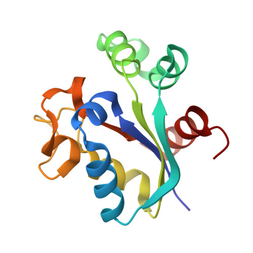
Structure of Mycobacterium Tuberculosis Nucleoside Diphosphate Kinase R80N Mutant in Complex with Citrate

Georgescauld, F.Moynie, L.Habersetzer, J.Dautant, A.

(2014) Acta Crystallogr D Biol Crystallogr 70: 40

  • DOI: https://doi.org/10.1107/S2053230X13034134
  • Primary Citation of Related Structures:  
    4ANE

  • PubMed Abstract: 

    The crystal structure of the wild-type nucleoside diphosphate kinase from Mycobacterium tuberculosis at 2.6 Å resolution revealed that the intersubunit salt bridge Arg80-Asp93 contributes to the thermal stability of the hexamer (Tm = 76°C). On mutating Asp93 to Asn to break the salt bridge, the thermal stability dramatically decreased by 27.6°C. Here, on mutating Arg80 to Asn, the thermal stability also significantly decreased by 8.0°C. In the X-ray structure of the R80N mutant solved at 1.9 Å resolution the salt bridge was replaced by intersubunit hydrogen bonds that contribute to the thermal stability of the hexamer. A citrate anion from the crystallization buffer was bound at the bottom of the nucleotide-binding site via electrostatic and hydrogen-bonding interactions with six conserved residues involved in nucleotide binding. Structural analysis shows that the citrate is present at the location of the nucleotide phosphate groups.


  • Organizational Affiliation

    Institut de Biochimie et de Génétique Cellulaires, UMR 5095, CNRS, 1 Rue Camille Saint-Saëns, 33077 Bordeaux, France.


Macromolecules
Find similar proteins by:  (by identity cutoff)  |  3D Structure
Entity ID: 1
MoleculeChains Sequence LengthOrganismDetailsImage
NUCLEOSIDE DIPHOSPHATE KINASE
A, B, C, D, E
A, B, C, D, E, F
136Mycobacterium tuberculosisMutation(s): 1 
EC: 2.7.4.6
UniProt
Find proteins for P9WJH7 (Mycobacterium tuberculosis (strain ATCC 25618 / H37Rv))
Explore P9WJH7 
Go to UniProtKB:  P9WJH7
Entity Groups  
Sequence Clusters30% Identity50% Identity70% Identity90% Identity95% Identity100% Identity
UniProt GroupP9WJH7
Sequence Annotations
Expand
  • Reference Sequence
Experimental Data & Validation

Experimental Data

  • Method: X-RAY DIFFRACTION
  • Resolution: 1.90 Å
  • R-Value Free: 0.235 
  • R-Value Work: 0.184 
  • R-Value Observed: 0.187 
  • Space Group: I 1 2 1
Unit Cell:
Length ( Å )Angle ( ˚ )
a = 68.82α = 90
b = 113.34β = 106.4
c = 107.561γ = 90
Software Package:
Software NamePurpose
PHENIXrefinement
MOSFLMdata reduction
SCALAdata scaling
MOLREPphasing

Structure Validation

View Full Validation Report


Entry History 

Deposition Data

Revision History  (Full details and data files)

  • Version 1.0: 2013-03-13
    Type: Initial release
  • Version 1.1: 2013-04-03
    Changes: Database references
  • Version 1.2: 2014-01-15
    Changes: Database references
  • Version 1.3: 2014-01-22
    Changes: Database references
  • Version 1.4: 2023-12-20
    Changes: Data collection, Database references, Derived calculations, Other, Refinement description