4AIV

Crystal Structure of putative NADH-dependent nitrite reductase small subunit from Mycobacterium tuberculosis


Experimental Data Snapshot

  • Method: X-RAY DIFFRACTION
  • Resolution: 2.00 Å
  • R-Value Free: 0.245 
  • R-Value Work: 0.194 
  • R-Value Observed: 0.197 

wwPDB Validation   3D Report Full Report


This is version 1.3 of the entry. See complete history


Literature

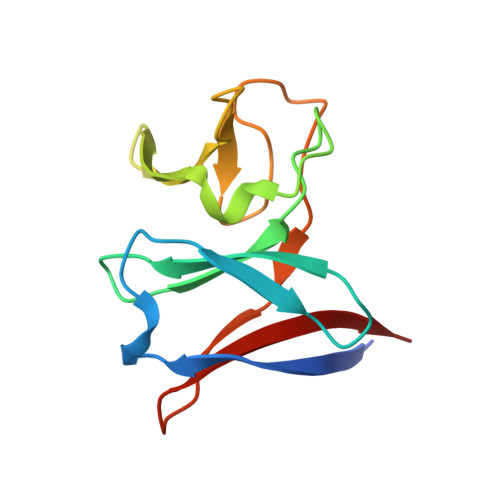
Crystal Structure of Nird, the Small Subunit of the Nitrite Reductase Nirbd from Mycobacterium Tuberculosis, at 2.0 Angstrom Resolution

Izumi, A.Schnell, R.Schneider, G.

(2012) Proteins 80: 2799

  • DOI: https://doi.org/10.1002/prot.24177
  • Primary Citation of Related Structures:  
    4AIV

  • PubMed Abstract: 

    NirD is part of the nitrite reductase complex NirBD that catalyses the reduction of nitrite to NH(3) in nitrate assimilation and anaerobic respiration. The crystal structure analysis of NirD from Mycobacterium tuberculosis shows a double β-sandwich fold. NirD is related in three-dimensional structure and sequence to the Rieske proteins; however, it does not contain any Fe-S cluster or other cofactors that might be involved in electron transfer. A cysteine residue at the protein surface, conserved in NirD homologues lacking the iron-sulfur cluster might be important for the interaction with NirB and possibly stabilize one of the Fe-S centers in this subunit.


  • Organizational Affiliation

    Department of Medical Biochemistry and Biophysics, Karolinska Institutet, S-17177 Stockholm, Sweden.


Macromolecules
Find similar proteins by:  (by identity cutoff)  |  3D Structure
Entity ID: 1
MoleculeChains Sequence LengthOrganismDetailsImage
PROBABLE NITRITE REDUCTASE [NAD(P)H] SMALL SUBUNIT NIRD119Mycobacterium tuberculosis H37RvMutation(s): 0 
EC: 1.6.6.4
UniProt
Find proteins for O53675 (Mycobacterium tuberculosis (strain ATCC 25618 / H37Rv))
Explore O53675 
Go to UniProtKB:  O53675
Entity Groups  
Sequence Clusters30% Identity50% Identity70% Identity90% Identity95% Identity100% Identity
UniProt GroupO53675
Sequence Annotations
Expand
  • Reference Sequence
Small Molecules
Ligands 1 Unique
IDChains Name / Formula / InChI Key2D Diagram3D Interactions
GOL
Query on GOL

Download Ideal Coordinates CCD File 
B [auth A]GLYCEROL
C3 H8 O3
PEDCQBHIVMGVHV-UHFFFAOYSA-N
Experimental Data & Validation

Experimental Data

  • Method: X-RAY DIFFRACTION
  • Resolution: 2.00 Å
  • R-Value Free: 0.245 
  • R-Value Work: 0.194 
  • R-Value Observed: 0.197 
  • Space Group: P 41 21 2
Unit Cell:
Length ( Å )Angle ( ˚ )
a = 35.104α = 90
b = 35.104β = 90
c = 180.392γ = 90
Software Package:
Software NamePurpose
PHENIXrefinement
XDSdata reduction
SCALAdata scaling
MOLREPphasing

Structure Validation

View Full Validation Report


Entry History 

Deposition Data

Revision History  (Full details and data files)

  • Version 1.0: 2012-09-12
    Type: Initial release
  • Version 1.1: 2012-09-26
    Changes: Database references
  • Version 1.2: 2012-11-14
    Changes: Database references
  • Version 1.3: 2023-12-20
    Changes: Data collection, Database references, Derived calculations, Other, Refinement description