


4AFA
Crystal Structure of subtype-switched Epithelial Adhesin 1 to 2 A domain (Epa1to2A) from Candida glabrata in complex with glycerol
- PDB DOI: https://doi.org/10.2210/pdb4AFA/pdb
- Classification: CELL ADHESION
- Organism(s): Nakaseomyces glabratus
- Expression System: Escherichia coli
- Mutation(s): Yes
- Deposited: 2012-01-18 Released: 2012-10-17
Experimental Data Snapshot
- Method: X-RAY DIFFRACTION
- Resolution: 2.05 Å
- R-Value Free: 0.239
- R-Value Work: 0.203
- R-Value Observed: 0.204
wwPDB Validation 3D Report Full Report

This is version 2.0 of the entry. See complete history.

Macromolecules
Find similar proteins by:
(by identity cutoff) | 3D Structure
Entity ID: 1 | |||||
|---|---|---|---|---|---|
| Molecule | Chains | Sequence Length | Organism | Details | Image |
| EPA1P | 262 | Nakaseomyces glabratus | Mutation(s): 3 | 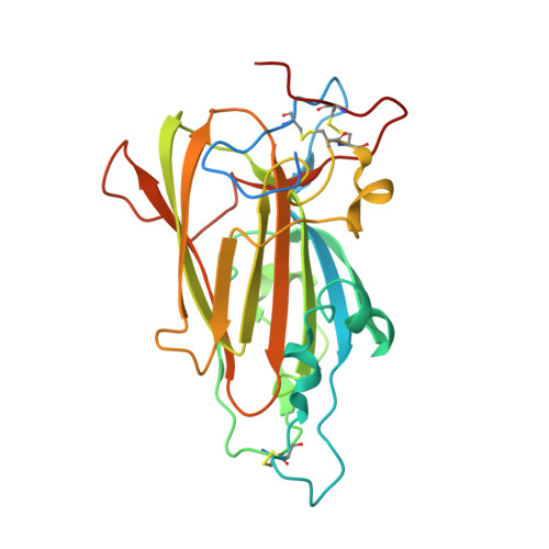 | |
UniProt | |||||
Find proteins for Q6VBJ0 (Candida glabrata) Explore Q6VBJ0 Go to UniProtKB: Q6VBJ0 | |||||
Entity Groups | |||||
| Sequence Clusters | 30% Identity50% Identity70% Identity90% Identity95% Identity100% Identity | ||||
| UniProt Group | Q6VBJ0 | ||||
Sequence AnnotationsExpand | |||||
| |||||
Oligosaccharides

Small Molecules
| Ligands 3 Unique | |||||
|---|---|---|---|---|---|
| ID | Chains | Name / Formula / InChI Key | 2D Diagram | 3D Interactions | |
| PEG Query on PEG | D [auth A] | DI(HYDROXYETHYL)ETHER C4 H10 O3 MTHSVFCYNBDYFN-UHFFFAOYSA-N |  | ||
| GOL Query on GOL | E [auth A], F [auth A], G [auth A] | GLYCEROL C3 H8 O3 PEDCQBHIVMGVHV-UHFFFAOYSA-N |  | ||
| CA Query on CA | C [auth A] | CALCIUM ION Ca BHPQYMZQTOCNFJ-UHFFFAOYSA-N |  | ||
Experimental Data & Validation
Experimental Data
- Method: X-RAY DIFFRACTION
- Resolution: 2.05 Å
- R-Value Free: 0.239
- R-Value Work: 0.203
- R-Value Observed: 0.204
- Space Group: C 2 2 21
Unit Cell:
| Length ( Å ) | Angle ( ˚ ) |
|---|---|
| a = 74.56 | α = 90 |
| b = 104.04 | β = 90 |
| c = 69.15 | γ = 90 |
| Software Name | Purpose |
|---|---|
| REFMAC | refinement |
| XDS | data reduction |
| SCALA | data scaling |
| XSCALE | data scaling |
| REFMAC | phasing |
Entry History
Deposition Data
- Released Date: 2012-10-17 Deposition Author(s): Maestre-Reyna, M., Diderrich, R., Veelders, M.S., Eulenburg, G., Kalugin, V., Brueckner, S., Keller, P., Rupp, S., Moesch, H.-U., Essen, L.-O.
Revision History (Full details and data files)
- Version 1.0: 2012-10-17
Type: Initial release - Version 1.1: 2012-10-31
Changes: Database references - Version 1.2: 2017-07-05
Changes: Data collection - Version 2.0: 2020-07-29
Type: Remediation
Reason: Carbohydrate remediation
Changes: Advisory, Atomic model, Data collection, Derived calculations, Other, Structure summary
