Crystal Structure of a Trypanosoma Brucei Metacaspase.
Mcluskey, K., Rudolf, J., Proto, W.R., Isaacs, N.W., Coombs, G.H., Moss, C.X., Mottram, J.C.(2012) Proc Natl Acad Sci U S A 109: 7469
- PubMed: 22529389
- DOI: https://doi.org/10.1073/pnas.1200885109
- Primary Citation of Related Structures:
4AF8, 4AFP, 4AFR, 4AFV - PubMed Abstract:
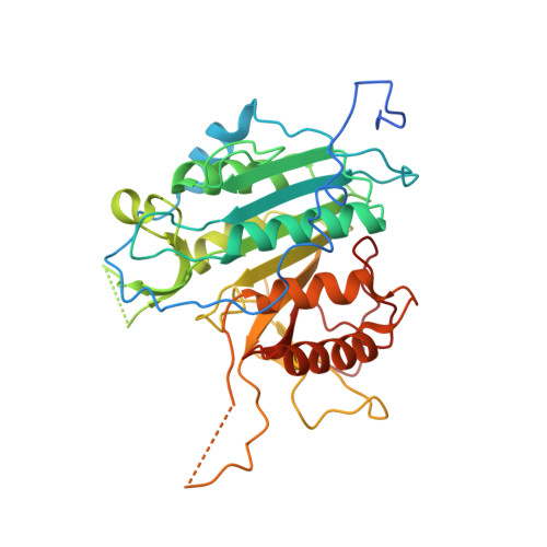
Metacaspases are distantly related caspase-family cysteine peptidases implicated in programmed cell death in plants and lower eukaryotes. They differ significantly from caspases because they are calcium-activated, arginine-specific peptidases that do not require processing or dimerization for activity. To elucidate the basis of these differences and to determine the impact they might have on the control of cell death pathways in lower eukaryotes, the previously undescribed crystal structure of a metacaspase, an inactive mutant of metacaspase 2 (MCA2) from Trypanosoma brucei, has been determined to a resolution of 1.4 Å. The structure comprises a core caspase fold, but with an unusual eight-stranded β-sheet that stabilizes the protein as a monomer. Essential aspartic acid residues, in the predicted S1 binding pocket, delineate the arginine-specific substrate specificity. In addition, MCA2 possesses an unusual N terminus, which encircles the protein and traverses the catalytic dyad, with Y31 acting as a gatekeeper residue. The calcium-binding site is defined by samarium coordinated by four aspartic acid residues, whereas calcium binding itself induces an allosteric conformational change that could stabilize the active site in a fashion analogous to subunit processing in caspases. Collectively, these data give insights into the mechanistic basis of substrate specificity and mode of activation of MCA2 and provide a detailed framework for understanding the role of metacaspases in cell death pathways of lower eukaryotes.
Organizational Affiliation:
Wellcome Trust Centre for Molecular Parasitology, Institute of Infection, Immunity, and Inflammation, College of Medical, Veterinary, and Life Sciences, University of Glasgow, Glasgow G12 8TA, United Kingdom. karen.mcluskey@glasgow.ac.uk