4ADY

Crystal structure of 26S proteasome subunit Rpn2


Experimental Data Snapshot

  • Method: X-RAY DIFFRACTION
  • Resolution: 2.70 Å
  • R-Value Free: 0.240 
  • R-Value Work: 0.183 
  • R-Value Observed: 0.184 

wwPDB Validation   3D Report Full Report


This is version 1.2 of the entry. See complete history


Literature

The Structure of the 26S Proteasome Subunit Rpn2 Reveals its Pc Repeat Domain as a Closed Toroid of Two Concentric Alpha-Helical Rings

He, J.Kulkarni, K.Da Fonseca, P.C.A.Krutauz, D.Glickman, M.H.Barford, D.Morris, E.P.

(2012) Structure 20: 513

  • DOI: https://doi.org/10.1016/j.str.2011.12.015
  • Primary Citation of Related Structures:  
    4ADY

  • PubMed Abstract: 

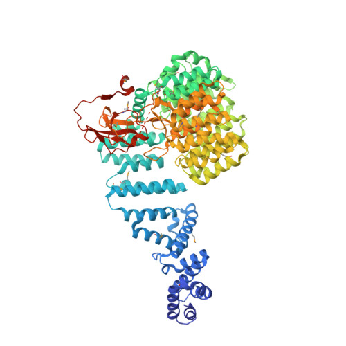
    The 26S proteasome proteolyses ubiquitylated proteins and is assembled from a 20S proteolytic core and two 19S regulatory particles (19S-RP). The 19S-RP scaffolding subunits Rpn1 and Rpn2 function to engage ubiquitin receptors. Rpn1 and Rpn2 are characterized by eleven tandem copies of a 35-40 amino acid repeat motif termed the proteasome/cyclosome (PC) repeat. Here, we reveal that the eleven PC repeats of Rpn2 form a closed toroidal structure incorporating two concentric rings of α helices encircling two axial α helices. A rod-like N-terminal domain consisting of 17 stacked α helices and a globular C-terminal domain emerge from one face of the toroid. Rpn13, an ubiquitin receptor, binds to the C-terminal 20 residues of Rpn2. Rpn1 adopts a similar conformation to Rpn2 but differs in the orientation of its rod-like N-terminal domain. These findings have implications for understanding how 19S-RPs recognize, unfold, and deliver ubiquitylated substrates to the 20S core.


  • Organizational Affiliation

    Division of Structural Biology, Institute of Cancer Research, Chester Beatty Laboratories, 237 Fulham Road, London SW3 6JB, United Kingdom.


Macromolecules
Find similar proteins by:  (by identity cutoff)  |  3D Structure
Entity ID: 1
MoleculeChains Sequence LengthOrganismDetailsImage
26S PROTEASOME REGULATORY SUBUNIT RPN2
A, B
963Saccharomyces cerevisiaeMutation(s): 0 
UniProt
Find proteins for P32565 (Saccharomyces cerevisiae (strain ATCC 204508 / S288c))
Explore P32565 
Go to UniProtKB:  P32565
Entity Groups  
Sequence Clusters30% Identity50% Identity70% Identity90% Identity95% Identity100% Identity
UniProt GroupP32565
Sequence Annotations
Expand
  • Reference Sequence
Small Molecules
Modified Residues  1 Unique
IDChains TypeFormula2D DiagramParent
MSE
Query on MSE
A, B
L-PEPTIDE LINKINGC5 H11 N O2 SeMET
Experimental Data & Validation

Experimental Data

  • Method: X-RAY DIFFRACTION
  • Resolution: 2.70 Å
  • R-Value Free: 0.240 
  • R-Value Work: 0.183 
  • R-Value Observed: 0.184 
  • Space Group: P 1 21 1
Unit Cell:
Length ( Å )Angle ( ˚ )
a = 73.96α = 90
b = 148.66β = 107.39
c = 114.03γ = 90
Software Package:
Software NamePurpose
MOSFLMdata reduction
SCALAdata scaling
SHELXphasing
SHARPphasing
PHENIXrefinement

Structure Validation

View Full Validation Report


Entry History 

Deposition Data

Revision History  (Full details and data files)

  • Version 1.0: 2012-03-14
    Type: Initial release
  • Version 1.1: 2012-03-21
    Changes: Other
  • Version 1.2: 2019-05-08
    Changes: Data collection, Derived calculations, Experimental preparation, Other