Hydroxamic Acids as Potent Inhibitors of Fe(II) and Mn(II) E. Coli Methionine Aminopeptidase: Biological Activities and X-Ray Structures of Oxazole Hydroxamate-Ecmetap-Mn Complexes.
Huguet, F., Melet, A., Alves De Sousa, R., Lieutaud, A., Chevalier, J., Maigre, L., Deschamps, P., Tomas, A., Leulliot, N., Pages, J.M., Artaud, I.(2012) ChemMedChem 7: 1020
- PubMed: 22489069
- DOI: https://doi.org/10.1002/cmdc.201200076
- Primary Citation of Related Structures:
4A6V, 4A6W - PubMed Abstract:
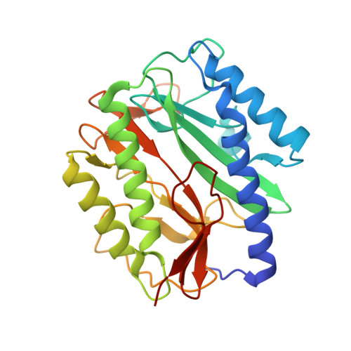
New series of acids and hydroxamic acids linked to five-membered heterocycles including furan, oxazole, 1,2,4- or 1,3,4-oxadiazole, and imidazole were synthesized and tested as inhibitors against the Fe(II) , Co(II) , and Mn(II) forms of E. coli methionine aminopeptidase (MetAP) and as antibacterial agents against wild-type and acrAB E. coli strains. 2-Aryloxazol-4-ylcarboxylic acids appeared as potent and selective inhibitors of the Co(II) MetAP form, with IC(50) values in the micromolar range, whereas 5-aryloxazol-2-ylcarboxylic acid regioisomers and 5-aryl-1,2,4-oxadiazol-3-ylcarboxylic acids were shown to be inefficient against all forms of EcMetAP. Regardless of the heterocycle, all the hydroxamic acids are highly potent inhibitors and are selective for the Mn(II) and Fe(II) forms, with IC(50) values between 1 and 2 μM. One indole hydroxamic acid that we previously reported as a potent inhibitor of E. coli peptide deformylase also demonstrated efficiency against EcMetAP. To gain insight into the positioning of the oxazole heterocycle with reversed substitutions at positions 2 and 5, X-ray crystal structures of EcMetAP-Mn complexed with two such oxazole hydroxamic acids were solved. Irrespective of the [metal]/[apo-MetAP] ratio, the active site consistently contains a dinuclear manganese center, with the hydroxamate as bridging ligand. Asp 97, which adopts a bidentate binding mode to the Mn2 site in the holoenzyme, is twisted in both structures toward the hydroxamate bridging ligand to favor the formation of a strong hydrogen bond. Most of the compounds show weak antibacterial activity against a wild-type E. coli strain. However, increased antibacterial activity was observed mainly for compounds with a 2-substituted phenyl group in the presence of the nonapeptide polymyxin B and phenylalanine-arginine-β-naphthylamide as permeabilizer and efflux pump blocker, respectively, which boost the intracellular uptake of the inhibitors.
Organizational Affiliation:
Laboratoire de Chimie et Biochimie Pharmacologiques et Toxicologiques, UMR 8601, CNRS Université Paris Descartes, 45 rue des Sts. Pères, 75270 Paris Cedex 06, France.