4A50

Crystal structure of human kinesin Eg5 in complex with 2-Amino-5-(3-methylphenyl)-5,5-diphenylpentanoic acid


Experimental Data Snapshot

  • Method: X-RAY DIFFRACTION
  • Resolution: 2.75 Å
  • R-Value Free: 0.243 
  • R-Value Work: 0.198 
  • R-Value Observed: 0.200 

wwPDB Validation   3D Report Full Report


Ligand Structure Quality Assessment 


This is version 1.2 of the entry. See complete history


Literature

Triphenylbutanamines: Kinesin Spindle Protein Inhibitors with in Vivo Antitumor Activity.

Wang, F.Good, J.A.D.Rath, O.Kaan, H.Y.K.Sutcliffe, O.B.Mackay, S.P.Kozielski, F.

(2012) J Med Chem 55: 1511

  • DOI: https://doi.org/10.1021/jm201195m
  • Primary Citation of Related Structures:  
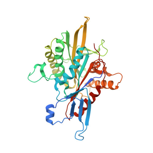
    4A50, 4A51

  • PubMed Abstract: 

    The human mitotic kinesin Eg5 represents a novel mitotic spindle target for cancer chemotherapy. We previously identified S-trityl-l-cysteine (STLC) and related analogues as selective potent inhibitors of Eg5. We herein report on the development of a series of 4,4,4-triphenylbutan-1-amine inhibitors derived from the STLC scaffold. This new generation systematically improves on potency: the most potent C-trityl analogues exhibit K(i)(app) ≤ 10 nM and GI(50) ≈ 50 nM, comparable to results from the phase II clinical benchmark ispinesib. Crystallographic studies reveal that they adopt the same overall binding configuration as S-trityl analogues at an allosteric site formed by loop L5 of Eg5. Evaluation of their druglike properties reveals favorable profiles for future development and, in the clinical candidate ispinesib, moderate hERG and CYP inhibition. One triphenylbutanamine analogue and ispinesib possess very good bioavailability (51% and 45%, respectively), with the former showing in vivo antitumor growth activity in nude mice xenograft studies.


  • Organizational Affiliation

    Molecular Motor Laboratory, The Beatson Institute for Cancer Research, Garscube Estate, Switchback Road, Glasgow G61 1BD, Scotland, UK. wangfang@moon.ibp.ac.cn


Macromolecules
Find similar proteins by:  (by identity cutoff)  |  3D Structure
Entity ID: 1
MoleculeChains Sequence LengthOrganismDetailsImage
KINESIN-LIKE PROTEIN KIF11368Homo sapiensMutation(s): 0 
UniProt & NIH Common Fund Data Resources
Find proteins for P52732 (Homo sapiens)
Explore P52732 
Go to UniProtKB:  P52732
PHAROS:  P52732
GTEx:  ENSG00000138160 
Entity Groups  
Sequence Clusters30% Identity50% Identity70% Identity90% Identity95% Identity100% Identity
UniProt GroupP52732
Sequence Annotations
Expand
  • Reference Sequence
Small Molecules
Ligands 4 Unique
IDChains Name / Formula / InChI Key2D Diagram3D Interactions
ADP
Query on ADP

Download Ideal Coordinates CCD File 
B [auth A]ADENOSINE-5'-DIPHOSPHATE
C10 H15 N5 O10 P2
XTWYTFMLZFPYCI-KQYNXXCUSA-N
DQ7
Query on DQ7

Download Ideal Coordinates CCD File 
E [auth A](2R)-2-AMINO-5-(3-METHYLPHENYL)-5,5-DIPHENYLPROPANOIC ACID
C24 H25 N O2
JXIGVUPYYZGRRZ-JOCHJYFZSA-N
DQ6
Query on DQ6

Download Ideal Coordinates CCD File 
D [auth A](2S)-2-AMINO-5-(3-METHYLPHENYL)-5,5-DIPHENYLPROPANOIC ACID
C24 H25 N O2
JXIGVUPYYZGRRZ-QFIPXVFZSA-N
MG
Query on MG

Download Ideal Coordinates CCD File 
C [auth A]MAGNESIUM ION
Mg
JLVVSXFLKOJNIY-UHFFFAOYSA-N
Binding Affinity Annotations 
IDSourceBinding Affinity
DQ7 BindingDB:  4A50 Ki: min: 5.1, max: 12.2 (nM) from 6 assay(s)
EC50: 73 (nM) from 1 assay(s)
DQ6 BindingDB:  4A50 Ki: min: 11.1, max: 12.8 (nM) from 2 assay(s)
Experimental Data & Validation

Experimental Data

  • Method: X-RAY DIFFRACTION
  • Resolution: 2.75 Å
  • R-Value Free: 0.243 
  • R-Value Work: 0.198 
  • R-Value Observed: 0.200 
  • Space Group: I 21 3
Unit Cell:
Length ( Å )Angle ( ˚ )
a = 158.54α = 90
b = 158.54β = 90
c = 158.54γ = 90
Software Package:
Software NamePurpose
REFMACrefinement
iMOSFLMdata reduction
SCALAdata scaling
REFMACphasing

Structure Validation

View Full Validation Report


Ligand Structure Quality Assessment 


Entry History 

Deposition Data

Revision History  (Full details and data files)

  • Version 1.0: 2012-10-31
    Type: Initial release
  • Version 1.1: 2013-02-27
    Changes: Database references, Other, Structure summary
  • Version 1.2: 2023-12-20
    Changes: Data collection, Database references, Derived calculations, Other, Refinement description