4A2L

Structure of the periplasmic domain of the heparin and heparan sulphate sensing hybrid two component system BT4663 in apo and ligand bound forms


Experimental Data Snapshot

  • Method: X-RAY DIFFRACTION
  • Resolution: 2.60 Å
  • R-Value Free: 0.231 
  • R-Value Work: 0.186 
  • R-Value Observed: 0.188 

wwPDB Validation   3D Report Full Report


This is version 1.2 of the entry. See complete history


Literature

A Scissor Blade-Like Closing Mechanism Implicated in Transmembrane Signaling in a Bacteroides Hybrid Two-Component System.

Lowe, E.C.Basle, A.Czjzek, M.Firbank, S.J.Bolam, D.N.

(2012) Proc Natl Acad Sci U S A 109: 7298

  • DOI: https://doi.org/10.1073/pnas.1200479109
  • Primary Citation of Related Structures:  
    4A2L, 4A2M

  • PubMed Abstract: 

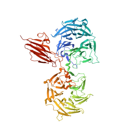
    Signaling across the membrane in response to extracellular stimuli is essential for survival of all cells. In bacteria, responses to environmental changes are predominantly mediated by two-component systems, which are typically composed of a membrane-spanning sensor histidine kinase and a cytoplasmic response regulator. In the human gut symbiont Bacteroides thetaiotaomicron, hybrid two-component systems are a key part of the bacterium's ability to sense and degrade complex carbohydrates in the gut. Here, we identify the activating ligand of the hybrid two-component system, BT4663, which controls heparin and heparan sulfate acquisition and degradation in this prominent gut microbe, and report the crystal structure of the extracellular sensor domain in both apo and ligand-bound forms. Current models for signal transduction across the membrane involve either a piston-like or rotational displacement of the transmembrane helices to modulate activity of the linked cytoplasmic kinases. The structures of the BT4663 sensor domain reveal a significant conformational change in the homodimer on ligand binding, which results in a scissor-like closing of the C-termini of each protomer. We propose this movement activates the attached intracellular kinase domains and represents an allosteric mechanism for bacterial transmembrane signaling distinct from previously described models, thus expanding our understanding of signal transduction across the membrane, a fundamental requirement in many important biological processes.


  • Organizational Affiliation

    Institute for Cell and Molecular Biosciences, Newcastle University, The Medical School, Newcastle upon Tyne NE2 4HH, United Kingdom.


Macromolecules
Find similar proteins by:  (by identity cutoff)  |  3D Structure
Entity ID: 1
MoleculeChains Sequence LengthOrganismDetailsImage
TWO-COMPONENT SYSTEM SENSOR HISTIDINE KINASE/RESPONSE
A, B, C, D, E
A, B, C, D, E, F
795Bacteroides thetaiotaomicron VPI-5482Mutation(s): 0 
UniProt
Find proteins for Q89YR8 (Bacteroides thetaiotaomicron (strain ATCC 29148 / DSM 2079 / JCM 5827 / CCUG 10774 / NCTC 10582 / VPI-5482 / E50))
Explore Q89YR8 
Go to UniProtKB:  Q89YR8
Entity Groups  
Sequence Clusters30% Identity50% Identity70% Identity90% Identity95% Identity100% Identity
UniProt GroupQ89YR8
Sequence Annotations
Expand
  • Reference Sequence
Small Molecules
Ligands 7 Unique
IDChains Name / Formula / InChI Key2D Diagram3D Interactions
2PE
Query on 2PE

Download Ideal Coordinates CCD File 
AA [auth D]NONAETHYLENE GLYCOL
C18 H38 O10
YZUUTMGDONTGTN-UHFFFAOYSA-N
MES
Query on MES

Download Ideal Coordinates CCD File 
CA [auth D],
GA [auth E],
P [auth B]
2-(N-MORPHOLINO)-ETHANESULFONIC ACID
C6 H13 N O4 S
SXGZJKUKBWWHRA-UHFFFAOYSA-N
PG4
Query on PG4

Download Ideal Coordinates CCD File 
L [auth B]TETRAETHYLENE GLYCOL
C8 H18 O5
UWHCKJMYHZGTIT-UHFFFAOYSA-N
PGE
Query on PGE

Download Ideal Coordinates CCD File 
EA [auth E],
G [auth A],
H [auth A],
HA [auth F],
M [auth B]
TRIETHYLENE GLYCOL
C6 H14 O4
ZIBGPFATKBEMQZ-UHFFFAOYSA-N
OAA
Query on OAA

Download Ideal Coordinates CCD File 
FA [auth E],
U [auth C],
V [auth C]
OXALOACETATE ION
C4 H3 O5
KHPXUQMNIQBQEV-UHFFFAOYSA-M
PEG
Query on PEG

Download Ideal Coordinates CCD File 
BA [auth D]
DA [auth E]
IA [auth F]
J [auth A]
K [auth B]
BA [auth D],
DA [auth E],
IA [auth F],
J [auth A],
K [auth B],
Q [auth C],
R [auth C],
T [auth C],
W [auth D],
X [auth D],
Y [auth D]
DI(HYDROXYETHYL)ETHER
C4 H10 O3
MTHSVFCYNBDYFN-UHFFFAOYSA-N
EDO
Query on EDO

Download Ideal Coordinates CCD File 
I [auth A],
N [auth B],
O [auth B],
S [auth C],
Z [auth D]
1,2-ETHANEDIOL
C2 H6 O2
LYCAIKOWRPUZTN-UHFFFAOYSA-N
Experimental Data & Validation

Experimental Data

  • Method: X-RAY DIFFRACTION
  • Resolution: 2.60 Å
  • R-Value Free: 0.231 
  • R-Value Work: 0.186 
  • R-Value Observed: 0.188 
  • Space Group: P 3 2 1
Unit Cell:
Length ( Å )Angle ( ˚ )
a = 342.468α = 90
b = 342.468β = 90
c = 97.852γ = 120
Software Package:
Software NamePurpose
REFMACrefinement
iMOSFLMdata reduction
SCALAdata scaling
SHELXCDEphasing

Structure Validation

View Full Validation Report


Entry History 

Deposition Data

Revision History  (Full details and data files)

  • Version 1.0: 2012-05-02
    Type: Initial release
  • Version 1.1: 2012-05-23
    Changes: Other
  • Version 1.2: 2014-11-19
    Changes: Data collection