4A03

Crystal Structure of Mycobacterium tuberculosis DXR in complex with the antibiotic FR900098 and cofactor NADPH


Experimental Data Snapshot

  • Method: X-RAY DIFFRACTION
  • Resolution: 1.65 Å
  • R-Value Free: 0.216 
  • R-Value Work: 0.189 
  • R-Value Observed: 0.191 

wwPDB Validation   3D Report Full Report


Ligand Structure Quality Assessment 


This is version 1.7 of the entry. See complete history


Literature

Structural Studies on Mycobacterium Tuberculosis Dxr in Complex with the Antibiotic Fr-900098.

Bjorkelid, C.Bergfors, T.Unge, T.Mowbray, S.L.Jones, T.A.

(2012) Acta Crystallogr D Biol Crystallogr 68: 134

  • DOI: https://doi.org/10.1107/S0907444911052231
  • Primary Citation of Related Structures:  
    4A03

  • PubMed Abstract: 

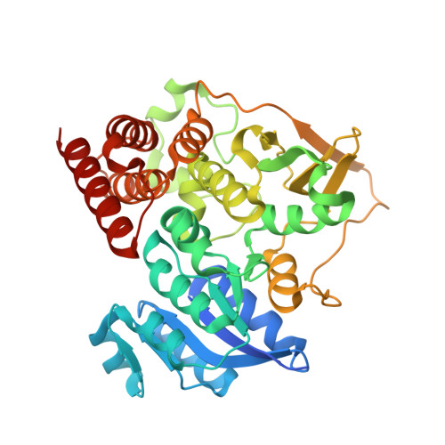
    A number of pathogens, including the causative agents of tuberculosis and malaria, synthesize the essential isoprenoid precursor isopentenyl diphosphate via the 2-C-methyl-D-erythritol 4-phosphate (MEP) pathway rather than the classical mevalonate pathway that is found in humans. As part of a structure-based drug-discovery program against tuberculosis, DXR, the enzyme that carries out the second step in the MEP pathway, has been investigated. This enzyme is the target for the antibiotic fosmidomycin and its active acetyl derivative FR-900098. The structure of DXR from Mycobacterium tuberculosis in complex with FR-900098, manganese and the NADPH cofactor has been solved and refined. This is a new crystal form that diffracts to a higher resolution than any other DXR complex reported to date. Comparisons with other ternary complexes show that the conformation is that of the enzyme in an active state: the active-site flap is well defined and the cofactor-binding domain has a conformation that brings the NADPH into the active site in a manner suitable for catalysis. The substrate-binding site is highly conserved in a number of pathogens that use this pathway, so any new inhibitor that is designed for the M. tuberculosis enzyme is likely to exhibit broad-spectrum activity.


  • Organizational Affiliation

    Department of Cell and Molecular Biology, Uppsala University, Biomedical Center, Box 596, SE-751 24 Uppsala, Sweden.


Macromolecules
Find similar proteins by:  (by identity cutoff)  |  3D Structure
Entity ID: 1
MoleculeChains Sequence LengthOrganismDetailsImage
1-DEOXY-D-XYLULOSE 5-PHOSPHATE REDUCTOISOMERASE
A, B
398Mycobacterium tuberculosis H37RvMutation(s): 0 
EC: 1.1.1.267
UniProt
Find proteins for P9WNS1 (Mycobacterium tuberculosis (strain ATCC 25618 / H37Rv))
Explore P9WNS1 
Go to UniProtKB:  P9WNS1
Entity Groups  
Sequence Clusters30% Identity50% Identity70% Identity90% Identity95% Identity100% Identity
UniProt GroupP9WNS1
Sequence Annotations
Expand
  • Reference Sequence
Small Molecules
Ligands 4 Unique
IDChains Name / Formula / InChI Key2D Diagram3D Interactions
NDP
Query on NDP

Download Ideal Coordinates CCD File 
E [auth A],
I [auth B]
NADPH DIHYDRO-NICOTINAMIDE-ADENINE-DINUCLEOTIDE PHOSPHATE
C21 H30 N7 O17 P3
ACFIXJIJDZMPPO-NNYOXOHSSA-N
F98
Query on F98

Download Ideal Coordinates CCD File 
D [auth A],
H [auth B]
3-[ethanoyl(hydroxy)amino]propylphosphonic acid
C5 H12 N O5 P
PKMNDDZSIHLLLI-UHFFFAOYSA-N
GOL
Query on GOL

Download Ideal Coordinates CCD File 
F [auth A]GLYCEROL
C3 H8 O3
PEDCQBHIVMGVHV-UHFFFAOYSA-N
MN
Query on MN

Download Ideal Coordinates CCD File 
C [auth A],
G [auth B]
MANGANESE (II) ION
Mn
WAEMQWOKJMHJLA-UHFFFAOYSA-N
Binding Affinity Annotations 
IDSourceBinding Affinity
F98 BindingDB:  4A03 Ki: 130 (nM) from 1 assay(s)
IC50: min: 160, max: 2390 (nM) from 2 assay(s)
Binding MOAD:  4A03 IC50: 160 (nM) from 1 assay(s)
Experimental Data & Validation

Experimental Data

  • Method: X-RAY DIFFRACTION
  • Resolution: 1.65 Å
  • R-Value Free: 0.216 
  • R-Value Work: 0.189 
  • R-Value Observed: 0.191 
  • Space Group: P 43 21 2
Unit Cell:
Length ( Å )Angle ( ˚ )
a = 110.51α = 90
b = 110.51β = 90
c = 137γ = 90
Software Package:
Software NamePurpose
REFMACrefinement
MOSFLMdata reduction
SCALAdata scaling
PHASERphasing

Structure Validation

View Full Validation Report


Ligand Structure Quality Assessment 


Entry History 

Deposition Data

Revision History  (Full details and data files)

  • Version 1.0: 2012-01-18
    Type: Initial release
  • Version 1.1: 2012-01-25
    Changes: Other
  • Version 1.2: 2012-03-28
    Changes: Other
  • Version 1.3: 2012-06-06
    Changes: Other
  • Version 1.4: 2019-03-06
    Changes: Data collection, Experimental preparation, Other
  • Version 1.5: 2019-05-15
    Changes: Data collection, Experimental preparation
  • Version 1.6: 2019-11-27
    Changes: Derived calculations, Other
  • Version 1.7: 2023-12-20
    Changes: Data collection, Database references, Derived calculations, Refinement description