4A01

Crystal Structure of the H-Translocating Pyrophosphatase


Experimental Data Snapshot

  • Method: X-RAY DIFFRACTION
  • Resolution: 2.35 Å
  • R-Value Free: 0.203 
  • R-Value Work: 0.168 
  • R-Value Observed: 0.170 

wwPDB Validation   3D Report Full Report


Ligand Structure Quality Assessment 


This is version 1.1 of the entry. See complete history


Literature

Crystal Structure of a Membrane Embedded H1-Translocating Pyrophosphatase

Lin, S.-M.Tsai, J.-Y.Hsiao, C.-D.Chiu, C.-L.Pan, R.-L.Sun, Y.-J.

(2012) Nature 484: 399

  • DOI: https://doi.org/10.1038/nature10963
  • Primary Citation of Related Structures:  
    4A01

  • PubMed Abstract: 

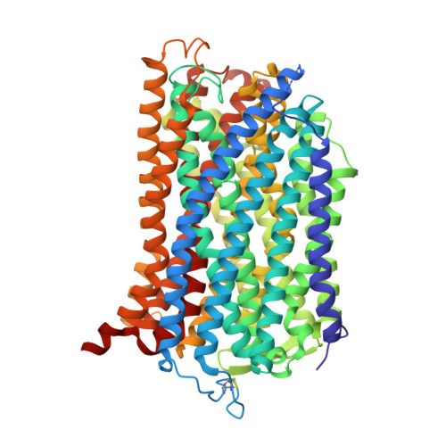
    H(+)-translocating pyrophosphatases (H(+)-PPases) are active proton transporters that establish a proton gradient across the endomembrane by means of pyrophosphate (PP(i)) hydrolysis. H(+)-PPases are found primarily as homodimers in the vacuolar membrane of plants and the plasma membrane of several protozoa and prokaryotes. The three-dimensional structure and detailed mechanisms underlying the enzymatic and proton translocation reactions of H(+)-PPases are unclear. Here we report the crystal structure of a Vigna radiata H(+)-PPase (VrH(+)-PPase) in complex with a non-hydrolysable substrate analogue, imidodiphosphate (IDP), at 2.35 Å resolution. Each VrH(+)-PPase subunit consists of an integral membrane domain formed by 16 transmembrane helices. IDP is bound in the cytosolic region of each subunit and trapped by numerous charged residues and five Mg(2+) ions. A previously undescribed proton translocation pathway is formed by six core transmembrane helices. Proton pumping can be initialized by PP(i) hydrolysis, and H(+) is then transported into the vacuolar lumen through a pathway consisting of Arg 242, Asp 294, Lys 742 and Glu 301. We propose a working model of the mechanism for the coupling between proton pumping and PP(i) hydrolysis by H(+)-PPases.


  • Organizational Affiliation

    Department of Life Science and Institute of Bioinformatics and Structural Biology, College of Life Science, National Tsing Hua University, Hsinchu 30013, Taiwan.


Macromolecules
Find similar proteins by:  (by identity cutoff)  |  3D Structure
Entity ID: 1
MoleculeChains Sequence LengthOrganismDetailsImage
PROTON PYROPHOSPHATASE
A, B
766Vigna radiataMutation(s): 0 
EC: 3.6.1.1
Membrane Entity: Yes 
UniProt
Find proteins for P21616 (Vigna radiata var. radiata)
Explore P21616 
Go to UniProtKB:  P21616
Entity Groups  
Sequence Clusters30% Identity50% Identity70% Identity90% Identity95% Identity100% Identity
UniProt GroupP21616
Sequence Annotations
Expand
  • Reference Sequence
Small Molecules
Ligands 4 Unique
IDChains Name / Formula / InChI Key2D Diagram3D Interactions
DMU
Query on DMU

Download Ideal Coordinates CCD File 
J [auth A]
K [auth A]
L [auth A]
M [auth A]
N [auth A]
J [auth A],
K [auth A],
L [auth A],
M [auth A],
N [auth A],
V [auth B],
W [auth B],
X [auth B],
Y [auth B]
DECYL-BETA-D-MALTOPYRANOSIDE
C22 H42 O11
WOQQAWHSKSSAGF-WXFJLFHKSA-N
2PN
Query on 2PN

Download Ideal Coordinates CCD File 
I [auth A],
U [auth B]
IMIDODIPHOSPHORIC ACID
H5 N O6 P2
GNGSOPFGGKKDQP-UHFFFAOYSA-N
K
Query on K

Download Ideal Coordinates CCD File 
H [auth A],
T [auth B]
POTASSIUM ION
K
NPYPAHLBTDXSSS-UHFFFAOYSA-N
MG
Query on MG

Download Ideal Coordinates CCD File 
C [auth A]
D [auth A]
E [auth A]
F [auth A]
G [auth A]
C [auth A],
D [auth A],
E [auth A],
F [auth A],
G [auth A],
O [auth B],
P [auth B],
Q [auth B],
R [auth B],
S [auth B]
MAGNESIUM ION
Mg
JLVVSXFLKOJNIY-UHFFFAOYSA-N
Experimental Data & Validation

Experimental Data

  • Method: X-RAY DIFFRACTION
  • Resolution: 2.35 Å
  • R-Value Free: 0.203 
  • R-Value Work: 0.168 
  • R-Value Observed: 0.170 
  • Space Group: C 1 2 1
Unit Cell:
Length ( Å )Angle ( ˚ )
a = 218.619α = 90
b = 88.241β = 125.53
c = 159.105γ = 90
Software Package:
Software NamePurpose
REFMACrefinement
HKL-2000data reduction
SCALEPACKdata scaling
PHENIXphasing

Structure Validation

View Full Validation Report


Ligand Structure Quality Assessment 


Entry History 

Deposition Data

Revision History  (Full details and data files)

  • Version 1.0: 2012-03-28
    Type: Initial release
  • Version 1.1: 2012-04-25
    Changes: Other