4ZLH

Structure of the LapB cytoplasmic domain at 2 angstroms


Experimental Data Snapshot

  • Method: X-RAY DIFFRACTION
  • Resolution: 2.00 Å
  • R-Value Free: 0.255 
  • R-Value Work: 0.203 
  • R-Value Observed: 0.206 

wwPDB Validation   3D Report Full Report


This is version 1.2 of the entry. See complete history


Literature

An Unexpected Duo: Rubredoxin Binds Nine TPR Motifs to Form LapB, an Essential Regulator of Lipopolysaccharide Synthesis.

Prince, C.Jia, Z.

(2015) Structure 23: 1500-1506

  • DOI: https://doi.org/10.1016/j.str.2015.06.011
  • Primary Citation of Related Structures:  
    4ZLH

  • PubMed Abstract: 

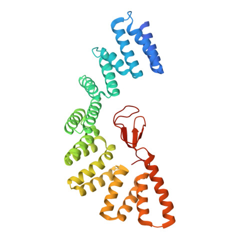
    Lipopolysaccharide (LPS) synthesis and export are essential pathways for bacterial growth, proliferation, and virulence. The essential protein LapB from Escherichia coli has recently been identified as a regulator of LPS synthesis. We have determined the crystal structure of LapB (without the N-terminal transmembrane helix) at 2 Å resolution using zinc single-wavelength anomalous diffraction phasing derived from a single bound zinc atom. This structure demonstrates the presence of nine tetratricopeptide repeats (TPR) motifs, including two TPR folds that were not predicted from sequence, and a rubredoxin-type metal binding domain. The rubredoxin domain is bound intimately to the TPR motifs, which has not been previously observed or predicted. Mutations in the rubredoxin/TPR interface inhibit in vivo cell growth, and in vitro studies indicate that these modifications cause local displacement of rubredoxin from its binding site without changing the secondary structure of LapB. LapB is the first reported structure to contain both a rubredoxin domain and TPR motifs.


  • Organizational Affiliation

    Department of Biomedical and Molecular Sciences, Queen's University, Kingston, ON K7L3N6, Canada.


Macromolecules
Find similar proteins by:  (by identity cutoff)  |  3D Structure
Entity ID: 1
MoleculeChains Sequence LengthOrganismDetailsImage
Lipopolysaccharide assembly protein B
A, B
339Escherichia coli O157:H7Mutation(s): 0 
Gene Names: lapByciMZ2526ECs1853
UniProt
Find proteins for P0AB60 (Escherichia coli O157:H7)
Explore P0AB60 
Go to UniProtKB:  P0AB60
Entity Groups  
Sequence Clusters30% Identity50% Identity70% Identity90% Identity95% Identity100% Identity
UniProt GroupP0AB60
Sequence Annotations
Expand
  • Reference Sequence
Experimental Data & Validation

Experimental Data

  • Method: X-RAY DIFFRACTION
  • Resolution: 2.00 Å
  • R-Value Free: 0.255 
  • R-Value Work: 0.203 
  • R-Value Observed: 0.206 
  • Space Group: P 1 21 1
Unit Cell:
Length ( Å )Angle ( ˚ )
a = 38.31α = 90
b = 152.25β = 93.91
c = 65.47γ = 90
Software Package:
Software NamePurpose
PHENIXrefinement
XDSdata reduction
XDSdata scaling
PHENIXphasing

Structure Validation

View Full Validation Report


Entry History & Funding Information

Deposition Data


Funding OrganizationLocationGrant Number
Canadian Institutes of Health Research (CIHR)Canada--

Revision History  (Full details and data files)

  • Version 1.0: 2015-11-11
    Type: Initial release
  • Version 1.1: 2020-01-08
    Changes: Author supporting evidence, Database references, Derived calculations
  • Version 1.2: 2024-03-06
    Changes: Data collection, Database references