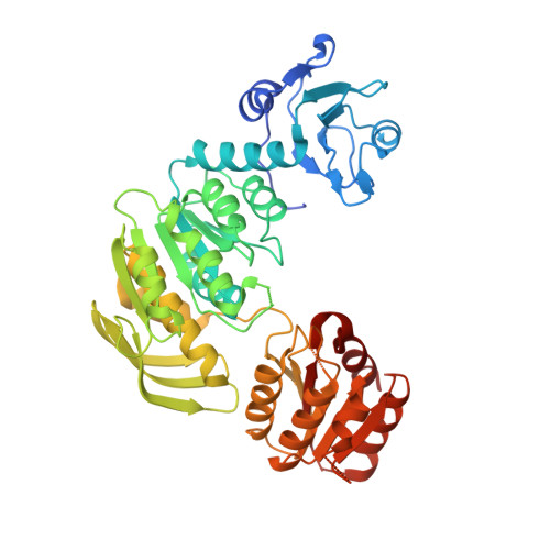
Structure of UDP-N-acetylmuramoylalanyl-D-glutamyl-2,6-diaminopimelate--D-alanyl-D-alanyl ligase from Acinetobacter baumannii
Seattle Structural Genomics Center for Infectious Disease (SSGCID), Abendroth, J., Clifton, M.C., Lorimer, D.D., Edwards, T.E.To be published.