4ZDX

Structure of OXA-51 beta-lactamase


Experimental Data Snapshot

  • Method: X-RAY DIFFRACTION
  • Resolution: 2.00 Å
  • R-Value Free: 0.216 
  • R-Value Work: 0.157 
  • R-Value Observed: 0.160 

wwPDB Validation   3D Report Full Report


Ligand Structure Quality Assessment 


This is version 1.1 of the entry. See complete history


Literature

Structural Basis for Enhancement of Carbapenemase Activity in the OXA-51 Family of Class D beta-Lactamases.

Smith, C.A.Antunes, N.T.Stewart, N.K.Frase, H.Toth, M.Kantardjieff, K.A.Vakulenko, S.

(2015) ACS Chem Biol 10: 1791-1796

  • DOI: https://doi.org/10.1021/acschembio.5b00090
  • Primary Citation of Related Structures:  
    4ZDX

  • PubMed Abstract: 

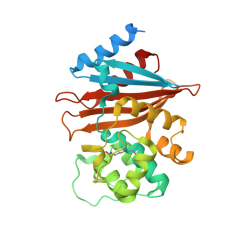
    Class D β-lactamases of Acinetobacter baumannii are enzymes of the utmost clinical importance, producing resistance to last resort carbapenem antibiotics. Although the OXA-51-like enzymes constitute the largest family of class D β-lactamases, they are poorly studied and their importance in conferring carbapenem resistance is controversial. We present the detailed microbiological, kinetic, and structural characterization of the eponymous OXA-51 β-lactamase. Kinetic studies show that OXA-51 has low catalytic efficiency for carbapenems, primarily due to the low affinity of the enzyme for these substrates. Structural studies demonstrate that this low affinity results from the obstruction of the enzyme active site by the side chain of Trp222, which presents a transient steric barrier to an incoming carbapenem substrate. The Trp222Met substitution relieves this steric hindrance and elevates the affinity of the mutant enzyme for carbapenems by 10-fold, significantly increasing the levels of resistance to these antibiotics. The ability of OXA-51 to evolve into a robust carbapenemase as the result of a single amino acid substitution may, in the near future, elevate the ubiquitous enzymes of the OXA-51 family to the status of the most deleterious A. baumannii carbapenemases, with dire clinical consequences.


  • Organizational Affiliation

    †Stanford Synchrotron Radiation Lightsource, Stanford University, Menlo Park, California 94025, United States.


Macromolecules
Find similar proteins by:  (by identity cutoff)  |  3D Structure
Entity ID: 1
MoleculeChains Sequence LengthOrganismDetailsImage
Beta-lactamase274Acinetobacter baumanniiMutation(s): 0 
UniProt
Find proteins for Q5QT35 (Acinetobacter baumannii)
Explore Q5QT35 
Go to UniProtKB:  Q5QT35
Entity Groups  
Sequence Clusters30% Identity50% Identity70% Identity90% Identity95% Identity100% Identity
UniProt GroupQ5QT35
Sequence Annotations
Expand
  • Reference Sequence
Small Molecules
Ligands 2 Unique
IDChains Name / Formula / InChI Key2D Diagram3D Interactions
TOE
Query on TOE

Download Ideal Coordinates CCD File 
B [auth A]2-[2-(2-METHOXY-ETHOXY)-ETHOXY]-ETHOXYL
C7 H16 O4
JLGLQAWTXXGVEM-UHFFFAOYSA-N
GOL
Query on GOL

Download Ideal Coordinates CCD File 
C [auth A]GLYCEROL
C3 H8 O3
PEDCQBHIVMGVHV-UHFFFAOYSA-N
Modified Residues  1 Unique
IDChains TypeFormula2D DiagramParent
KCX
Query on KCX
A
L-PEPTIDE LINKINGC7 H14 N2 O4LYS
Experimental Data & Validation

Experimental Data

  • Method: X-RAY DIFFRACTION
  • Resolution: 2.00 Å
  • R-Value Free: 0.216 
  • R-Value Work: 0.157 
  • R-Value Observed: 0.160 
  • Space Group: I 4 2 2
Unit Cell:
Length ( Å )Angle ( ˚ )
a = 131.111α = 90
b = 131.111β = 90
c = 67.525γ = 90
Software Package:
Software NamePurpose
PHENIXrefinement
XDSdata reduction
Aimlessdata scaling
MOLREPphasing

Structure Validation

View Full Validation Report


Ligand Structure Quality Assessment 


Entry History 

Deposition Data

Revision History  (Full details and data files)

  • Version 1.0: 2015-06-17
    Type: Initial release
  • Version 1.1: 2015-09-02
    Changes: Database references