4YR6

Fab fragment of 5G6 in complex with epitope peptide


Experimental Data Snapshot

  • Method: X-RAY DIFFRACTION
  • Resolution: 2.38 Å
  • R-Value Free: 0.251 
  • R-Value Work: 0.211 
  • R-Value Observed: 0.213 

wwPDB Validation   3D Report Full Report


This is version 1.3 of the entry. See complete history


Literature

Structural basis for the specific inhibition of glycoprotein Ib alpha shedding by an inhibitory antibody.

Tao, Y.Zhang, X.Liang, X.Zang, J.Mo, X.Li, R.

(2016) Sci Rep 6: 24789-24789

  • DOI: https://doi.org/10.1038/srep24789
  • Primary Citation of Related Structures:  
    4YR6

  • PubMed Abstract: 

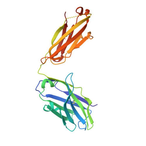
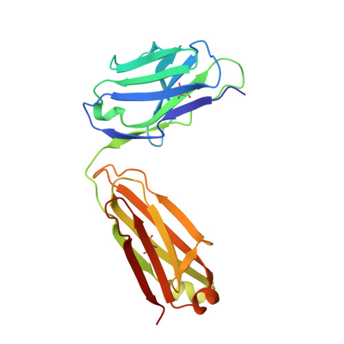
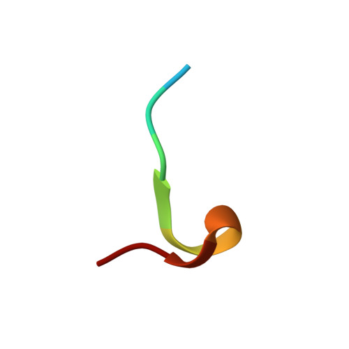
    Ectodomain shedding of glycoprotein (GP) Ibα is thought to mediate the clearance of activated, aged or damaged platelets. A monoclonal antibody, 5G6, has been developed recently to specifically bind to the GPIbα shedding cleavage site and to inhibit its shedding. However, the molecular mechanism underlying antigen recognition and inhibitory specificity is not clear. To elucidate the structural basis for 5G6 binding to GPIbα, we determined the crystal structure of 5G6 Fab fragment in complex with its epitope peptide KL10 (GPIbα residues 461-470, KLRGVLQGHL), to 2.4-Å resolution. Key residues in both 5G6 and KL10 were mutated to validate their effects in antibody binding by using isothermal titration calorimetry. The 5G6 Fab-KL10 peptide complex structure confirmed the direct association of 5G6 with its target GPIbα residues and elucidated the molecular basis underlying its binding specificity and high affinity. The similar binding properties of 5G6 Fab fragment to GPIbα on human platelets as those to KL10 suggests that such an interaction may not be affected by the plasma membrane or nearby GPIbβ. This structural information may facilitate further antibody optimization and humanization.


  • Organizational Affiliation

    Key Laboratory of Pediatric Hematology &Oncology Ministry of Health, Pediatric Translational Medicine Institute, Shanghai Children's Medical Center, Shanghai Jiao Tong University School of Medicine, Shanghai, China.


Macromolecules
Find similar proteins by:  (by identity cutoff)  |  3D Structure
Entity ID: 1
MoleculeChains Sequence LengthOrganismDetailsImage
heavy chain of 5G6
A, D
221Mus musculusMutation(s): 0 
Entity Groups  
Sequence Clusters30% Identity50% Identity70% Identity90% Identity95% Identity100% Identity
Sequence Annotations
Expand
  • Reference Sequence
Find similar proteins by:  (by identity cutoff)  |  3D Structure
Entity ID: 2
MoleculeChains Sequence LengthOrganismDetailsImage
light chain of 5G6
B, E
214Mus musculusMutation(s): 0 
Entity Groups  
Sequence Clusters30% Identity50% Identity70% Identity90% Identity95% Identity100% Identity
Sequence Annotations
Expand
  • Reference Sequence

Find similar proteins by:  Sequence   |   3D Structure  

Entity ID: 3
MoleculeChains Sequence LengthOrganismDetailsImage
ACE-LYS-LEU-ARG-GLY-VAL-LEU-GLN-GLY-HIS-LEU
C, F
11Homo sapiensMutation(s): 0 
UniProt & NIH Common Fund Data Resources
Find proteins for P07359 (Homo sapiens)
Explore P07359 
Go to UniProtKB:  P07359
PHAROS:  P07359
GTEx:  ENSG00000185245 
Entity Groups  
Sequence Clusters30% Identity50% Identity70% Identity90% Identity95% Identity100% Identity
UniProt GroupP07359
Sequence Annotations
Expand
  • Reference Sequence
Experimental Data & Validation

Experimental Data

  • Method: X-RAY DIFFRACTION
  • Resolution: 2.38 Å
  • R-Value Free: 0.251 
  • R-Value Work: 0.211 
  • R-Value Observed: 0.213 
  • Space Group: C 2 2 21
Unit Cell:
Length ( Å )Angle ( ˚ )
a = 101.9α = 90
b = 147.06β = 90
c = 194.95γ = 90
Software Package:
Software NamePurpose
PHENIXrefinement
PHENIXmodel building
HKL-2000data scaling

Structure Validation

View Full Validation Report


Entry History & Funding Information

Deposition Data


Funding OrganizationLocationGrant Number
China Natural Science FundChina81100346
Shanghai Xinyouqing ProgramChinaXYQ2013069

Revision History  (Full details and data files)

  • Version 1.0: 2016-03-09
    Type: Initial release
  • Version 1.1: 2017-04-05
    Changes: Source and taxonomy
  • Version 1.2: 2020-05-13
    Changes: Database references
  • Version 1.3: 2023-11-08
    Changes: Data collection, Database references, Refinement description