4YNG

Twinned pyruvate kinase from E. coli in the T-state


Experimental Data Snapshot

  • Method: X-RAY DIFFRACTION
  • Resolution: 2.28 Å
  • R-Value Free: 0.244 
  • R-Value Work: 0.217 
  • R-Value Observed: 0.219 

wwPDB Validation   3D Report Full Report


This is version 1.5 of the entry. See complete history


Literature

Grappling with anisotropic data, pseudo-merohedral twinning and pseudo-translational noncrystallographic symmetry: a case study involving pyruvate kinase.

Donovan, K.A.Atkinson, S.C.Kessans, S.A.Peng, F.Cooper, T.F.Griffin, M.D.Jameson, G.B.Dobson, R.C.

(2016) Acta Crystallogr D Struct Biol 72: 512-519

  • DOI: https://doi.org/10.1107/S205979831600142X
  • Primary Citation of Related Structures:  
    4YNG

  • PubMed Abstract: 

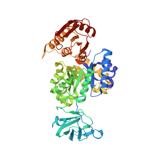
    Pyruvate kinase is a key regulatory enzyme involved in the glycolytic pathway. The crystal structure of Escherichia coli type I pyruvate kinase was first solved in 1995 at 2.5 Å resolution. However, the space group was ambiguous, being either primitive orthorhombic (P2(1)2(1)2(1)) or C-centred orthorhombic (C222(1)). Here, the structure determination and refinement of E. coli type I pyruvate kinase to 2.28 Å resolution are presented. Using the same crystallization conditions as reported previously, the enzyme was found to crystallize in space group P2(1). Determination of the space group was complicated owing to anisotropic data, pseudo-translational noncrystallographic symmetry and the pseudo-merohedrally twinned nature of the crystal, which was found to have very close to 50% twinning, leading to apparent orthorhombic symmetry and absences that were not inconsistent with P2(1)2(1)2(1). The unit cell contained two tetramers in the asymmetric unit (3720 residues) and, when compared with the orthorhombic structure, virtually all of the residues could be easily modelled into the density. Averaging of reflections into the lower symmetry space group with twinning provided tidier electron density that allowed ∼30 missing residues of the lid domain to be modelled for the first time. Moreover, residues in a flexible loop could be modelled and sulfate molecules are found in the allosteric binding domain, identifying the pocket that binds the allosteric activator fructose 1,6-bisphosphate in this isozyme for the first time. Lastly, we note the pedagogical benefits of difficult structures to emerging crystallographers.


  • Organizational Affiliation

    Biomolecular Interaction Centre and School of Biological Sciences, University of Canterbury, Private Bag 4800, Christchurch 8041, New Zealand.


Macromolecules
Find similar proteins by:  (by identity cutoff)  |  3D Structure
Entity ID: 1
MoleculeChains Sequence LengthOrganismDetailsImage
Pyruvate kinase I
A, B, C, D, E
A, B, C, D, E, F, G, H
470Escherichia coliMutation(s): 0 
Gene Names: pykFZ2704ECs2383
EC: 2.7.1.40
UniProt
Find proteins for P0AD62 (Escherichia coli O157:H7)
Explore P0AD62 
Go to UniProtKB:  P0AD62
Entity Groups  
Sequence Clusters30% Identity50% Identity70% Identity90% Identity95% Identity100% Identity
UniProt GroupP0AD62
Sequence Annotations
Expand
  • Reference Sequence
Small Molecules
Ligands 1 Unique
IDChains Name / Formula / InChI Key2D Diagram3D Interactions
SO4
Query on SO4

Download Ideal Coordinates CCD File 
I [auth A]
J [auth B]
K [auth B]
L [auth C]
M [auth D]
I [auth A],
J [auth B],
K [auth B],
L [auth C],
M [auth D],
N [auth D],
O [auth E],
P [auth F],
Q [auth F],
R [auth G],
S [auth H],
T [auth H]
SULFATE ION
O4 S
QAOWNCQODCNURD-UHFFFAOYSA-L
Experimental Data & Validation

Experimental Data

  • Method: X-RAY DIFFRACTION
  • Resolution: 2.28 Å
  • R-Value Free: 0.244 
  • R-Value Work: 0.217 
  • R-Value Observed: 0.219 
  • Space Group: P 1 21 1
Unit Cell:
Length ( Å )Angle ( ˚ )
a = 130.09α = 90
b = 74.596β = 90.14
c = 241.634γ = 90
Software Package:
Software NamePurpose
Aimlessdata scaling
PHASERphasing
PHENIXrefinement
PDB_EXTRACTdata extraction
REFMACrefinement
Cootmodel building

Structure Validation

View Full Validation Report


Entry History & Funding Information

Deposition Data


Funding OrganizationLocationGrant Number
Royal Society Marsden FundNew ZealandUOC1013
US Army Research LaboratoryNew ZealandW911NF-11-1-0481

Revision History  (Full details and data files)

  • Version 1.0: 2015-03-25
    Type: Initial release
  • Version 1.1: 2016-03-09
    Changes: Experimental preparation
  • Version 1.2: 2016-04-13
    Changes: Database references
  • Version 1.3: 2017-11-22
    Changes: Data collection, Derived calculations, Refinement description
  • Version 1.4: 2018-04-18
    Changes: Data collection, Database references
  • Version 1.5: 2024-02-28
    Changes: Data collection, Database references, Refinement description