4YLU

X-ray structure of MERS-CoV nsp5 protease bound with a non-covalent inhibitor


Experimental Data Snapshot

  • Method: X-RAY DIFFRACTION
  • Resolution: 2.10 Å
  • R-Value Free: 0.215 
  • R-Value Work: 0.159 
  • R-Value Observed: 0.161 

wwPDB Validation   3D Report Full Report


Ligand Structure Quality Assessment 


This is version 1.5 of the entry. See complete history


Literature

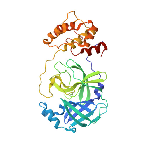
Ligand-induced Dimerization of Middle East Respiratory Syndrome (MERS) Coronavirus nsp5 Protease (3CLpro): IMPLICATIONS FOR nsp5 REGULATION AND THE DEVELOPMENT OF ANTIVIRALS.

Tomar, S.Johnston, M.L.St John, S.E.Osswald, H.L.Nyalapatla, P.R.Paul, L.N.Ghosh, A.K.Denison, M.R.Mesecar, A.D.

(2015) J Biol Chem 290: 19403-19422

  • DOI: https://doi.org/10.1074/jbc.M115.651463
  • Primary Citation of Related Structures:  
    4RSP, 4YLU

  • PubMed Abstract: 

    All coronaviruses, including the recently emerged Middle East respiratory syndrome coronavirus (MERS-CoV) from the β-CoV subgroup, require the proteolytic activity of the nsp5 protease (also known as 3C-like protease, 3CL(pro)) during virus replication, making it a high value target for the development of anti-coronavirus therapeutics. Kinetic studies indicate that in contrast to 3CL(pro) from other β-CoV 2c members, including HKU4 and HKU5, MERS-CoV 3CL(pro) is less efficient at processing a peptide substrate due to MERS-CoV 3CL(pro) being a weakly associated dimer. Conversely, HKU4, HKU5, and SARS-CoV 3CL(pro) enzymes are tightly associated dimers. Analytical ultracentrifugation studies support that MERS-CoV 3CL(pro) is a weakly associated dimer (Kd ∼52 μm) with a slow off-rate. Peptidomimetic inhibitors of MERS-CoV 3CL(pro) were synthesized and utilized in analytical ultracentrifugation experiments and demonstrate that MERS-CoV 3CL(pro) undergoes significant ligand-induced dimerization. Kinetic studies also revealed that designed reversible inhibitors act as activators at a low compound concentration as a result of induced dimerization. Primary sequence comparisons and x-ray structural analyses of two MERS-CoV 3CLpro and inhibitor complexes, determined to 1.6 Å, reveal remarkable structural similarity of the dimer interface with 3CL(pro) from HKU4-CoV and HKU5-CoV. Despite this structural similarity, substantial differences in the dimerization ability suggest that long range interactions by the nonconserved amino acids distant from the dimer interface may control MERS-CoV 3CL(pro) dimerization. Activation of MERS-CoV 3CL(pro) through ligand-induced dimerization appears to be unique within the genogroup 2c and may potentially increase the complexity in the development of MERS-CoV 3CL(pro) inhibitors as antiviral agents.


  • Organizational Affiliation

    From the Departments of Biological Sciences and.


Macromolecules
Find similar proteins by:  (by identity cutoff)  |  3D Structure
Entity ID: 1
MoleculeChains Sequence LengthOrganismDetailsImage
ORF1a protein
A, B, C, D
306Middle East respiratory syndrome-related coronavirusMutation(s): 0 
Gene Names: orf1ab
UniProt
Find proteins for V9TU12 (Middle East respiratory syndrome-related coronavirus)
Explore V9TU12 
Go to UniProtKB:  V9TU12
Entity Groups  
Sequence Clusters30% Identity50% Identity70% Identity90% Identity95% Identity100% Identity
UniProt GroupV9TU12
Sequence Annotations
Expand
  • Reference Sequence
Small Molecules
Ligands 2 Unique
IDChains Name / Formula / InChI Key2D Diagram3D Interactions
R30
Query on R30

Download Ideal Coordinates CCD File 
E [auth A],
H [auth B],
K [auth C],
M [auth D]
N-{4-[(1H-benzotriazol-1-ylacetyl)(thiophen-3-ylmethyl)amino]phenyl}propanamide
C22 H21 N5 O2 S
TWIVXCFEBRGEKY-UHFFFAOYSA-N
ACT
Query on ACT

Download Ideal Coordinates CCD File 
F [auth A]
G [auth A]
I [auth B]
J [auth B]
L [auth C]
F [auth A],
G [auth A],
I [auth B],
J [auth B],
L [auth C],
N [auth D]
ACETATE ION
C2 H3 O2
QTBSBXVTEAMEQO-UHFFFAOYSA-M
Experimental Data & Validation

Experimental Data

  • Method: X-RAY DIFFRACTION
  • Resolution: 2.10 Å
  • R-Value Free: 0.215 
  • R-Value Work: 0.159 
  • R-Value Observed: 0.161 
  • Space Group: P 1 21 1
Unit Cell:
Length ( Å )Angle ( ˚ )
a = 63.439α = 90
b = 114.93β = 90.89
c = 92.341γ = 90
Software Package:
Software NamePurpose
PHENIXrefinement
HKL-2000data collection
HKL-2000data scaling
PDB_EXTRACTdata extraction
HKL-2000data reduction
PHASERphasing

Structure Validation

View Full Validation Report


Ligand Structure Quality Assessment 


Entry History & Funding Information

Deposition Data


Funding OrganizationLocationGrant Number
National Institutes of Health/National Institute Of Allergy and Infectious Diseases (NIH/NIAID)United StatesAI08508 to ADM
National Institutes of Health/National Institute Of Allergy and Infectious Diseases (NIH/NIAID)United StatesAI026603 to ADM and MRD

Revision History  (Full details and data files)

  • Version 1.0: 2015-06-17
    Type: Initial release
  • Version 1.1: 2015-07-15
    Changes: Database references
  • Version 1.2: 2015-08-19
    Changes: Database references
  • Version 1.3: 2017-09-20
    Changes: Advisory, Author supporting evidence, Database references, Derived calculations
  • Version 1.4: 2019-12-11
    Changes: Author supporting evidence
  • Version 1.5: 2024-02-28
    Changes: Advisory, Data collection, Database references