


Structure of a putative phosphomethylpyrimidine kinase from Acinetobacter baumannii.
Abendroth, J., Lukacs, C.M., Lorimer, D.D., Edwards, T.E.To be published.
Experimental Data Snapshot
wwPDB Validation 3D Report Full Report

Entity ID: 1 | |||||
|---|---|---|---|---|---|
| Molecule | Chains | Sequence Length | Organism | Details | Image |
| Putative phosphomethylpyrimidine kinase | 263 | Acinetobacter baumannii IS-123 | Mutation(s): 0 Gene Names: ACINIS123_0279 | 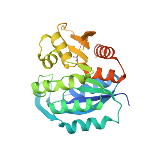 | |
UniProt | |||||
Find proteins for A0A0J9X285 (Acinetobacter baumannii IS-123) Explore A0A0J9X285 Go to UniProtKB: A0A0J9X285 | |||||
Entity Groups | |||||
| Sequence Clusters | 30% Identity50% Identity70% Identity90% Identity95% Identity100% Identity | ||||
| UniProt Group | A0A0J9X285 | ||||
Sequence AnnotationsExpand | |||||
| |||||
| Ligands 1 Unique | |||||
|---|---|---|---|---|---|
| ID | Chains | Name / Formula / InChI Key | 2D Diagram | 3D Interactions | |
| EDO Query on EDO | B [auth A], C [auth A] | 1,2-ETHANEDIOL C2 H6 O2 LYCAIKOWRPUZTN-UHFFFAOYSA-N |  | ||
| Length ( Å ) | Angle ( ˚ ) |
|---|---|
| a = 58.82 | α = 90 |
| b = 108.1 | β = 90 |
| c = 88.88 | γ = 90 |
| Software Name | Purpose |
|---|---|
| XDS | data scaling |
| BALBES | phasing |
| PHASER | phasing |
| PHENIX | refinement |
| PDB_EXTRACT | data extraction |
RCSB PDB (citation) is hosted by
RCSB PDB is a member of the

