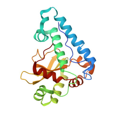
4YET

X-ray crystal structure of superoxide dismutase from Babesia bovis solved by Sulfur SAD


Experimental Data Snapshot

  • Method: X-RAY DIFFRACTION
  • Resolution: 1.75 Å
  • R-Value Free: 0.176 
  • R-Value Work: 0.143 
  • R-Value Observed: 0.145 

wwPDB Validation   3D Report Full Report


This is version 1.5 of the entry. See complete history


Literature

Iron superoxide dismutases in eukaryotic pathogens: new insights from Apicomplexa and Trypanosoma structures.

Phan, I.Q.Davies, D.R.Moretti, N.S.Shanmugam, D.Cestari, I.Anupama, A.Fairman, J.W.Edwards, T.E.Stuart, K.Schenkman, S.Myler, P.J.

(2015) Acta Crystallogr F Struct Biol Commun 71: 615-621

  • DOI: https://doi.org/10.1107/S2053230X15004185
  • Primary Citation of Related Structures:  
    4F2N, 4H3E, 4YET

  • PubMed Abstract: 

    Prior studies have highlighted the potential of superoxide dismutases as drug targets in eukaryotic pathogens. This report presents the structures of three iron-dependent superoxide dismutases (FeSODs) from Trypanosoma cruzi, Leishmania major and Babesia bovis. Comparison with existing structures from Plasmodium and other trypanosome isoforms shows a very conserved overall fold with subtle differences. In particular, structural data suggest that B. bovis FeSOD may display similar resistance to peroxynitrite-mediated inactivation via an intramolecular electron-transfer pathway as previously described in T. cruzi FeSOD isoform B, thus providing valuable information for structure-based drug design. Furthermore, lysine-acetylation results in T. cruzi indicate that acetylation occurs at a position close to that responsible for the regulation of acetylation-mediated activity in the human enzyme.


  • Organizational Affiliation

    Seattle Structural Genomics Center for Infectious Disease (SSGCID), USA.


Macromolecules
Find similar proteins by:  (by identity cutoff)  |  3D Structure
Entity ID: 1
MoleculeChains Sequence LengthOrganismDetailsImage
Superoxide dismutase
A, B
203Babesia bovisMutation(s): 0 
Gene Names: Fe-SODBBOV_IV011480
EC: 1.15.1.1
UniProt
Find proteins for O15904 (Babesia bovis)
Explore O15904 
Go to UniProtKB:  O15904
Entity Groups  
Sequence Clusters30% Identity50% Identity70% Identity90% Identity95% Identity100% Identity
UniProt GroupO15904
Sequence Annotations
Expand
  • Reference Sequence
Experimental Data & Validation

Experimental Data

  • Method: X-RAY DIFFRACTION
  • Resolution: 1.75 Å
  • R-Value Free: 0.176 
  • R-Value Work: 0.143 
  • R-Value Observed: 0.145 
  • Space Group: P 21 21 21
Unit Cell:
Length ( Å )Angle ( ˚ )
a = 55.75α = 90
b = 69.09β = 90
c = 114.81γ = 90
Software Package:
Software NamePurpose
XSCALEdata scaling
PHENIXrefinement
PDB_EXTRACTdata extraction
PHASERphasing

Structure Validation

View Full Validation Report


Entry History & Funding Information

Deposition Data


Funding OrganizationLocationGrant Number
National Institutes of Health/National Institute Of Allergy and Infectious Diseases (NIH/NIAID)United States--

Revision History  (Full details and data files)

  • Version 1.0: 2015-04-15
    Type: Initial release
  • Version 1.1: 2015-06-03
    Changes: Database references
  • Version 1.2: 2017-09-13
    Changes: Author supporting evidence, Database references, Derived calculations, Source and taxonomy
  • Version 1.3: 2017-11-22
    Changes: Refinement description
  • Version 1.4: 2019-12-11
    Changes: Author supporting evidence
  • Version 1.5: 2024-02-28
    Changes: Data collection, Database references, Derived calculations