4YDU

Crystal structure of E. coli YgjD-YeaZ heterodimer in complex with ADP


Experimental Data Snapshot

  • Method: X-RAY DIFFRACTION
  • Resolution: 2.33 Å
  • R-Value Free: 0.253 
  • R-Value Work: 0.202 
  • R-Value Observed: 0.205 

wwPDB Validation   3D Report Full Report


Ligand Structure Quality Assessment 


This is version 1.1 of the entry. See complete history


Literature

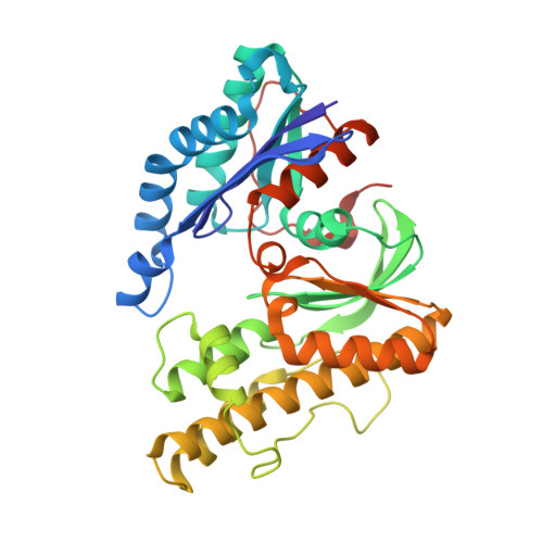
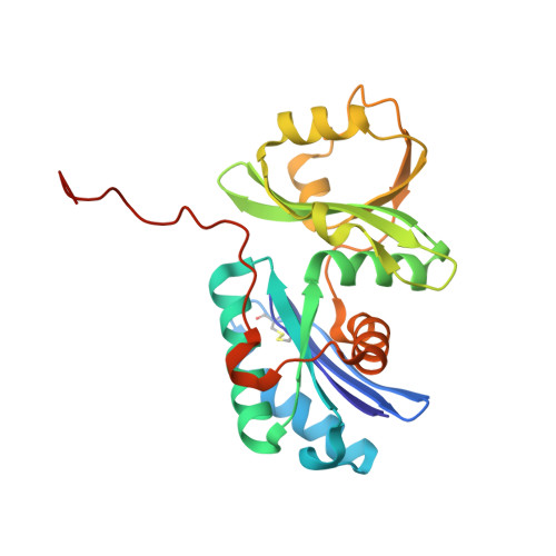
The ATP-mediated formation of the YgjD-YeaZ-YjeE complex is required for the biosynthesis of tRNA t6A in Escherichia coli.

Zhang, W.Collinet, B.Perrochia, L.Durand, D.Van Tilbeurgh, H.

(2015) Nucleic Acids Res 43: 1804-1817

  • DOI: https://doi.org/10.1093/nar/gku1397
  • Primary Citation of Related Structures:  
    4WQ4, 4WQ5, 4YDU

  • PubMed Abstract: 

    The essential and universal N(6)-threonylcarbamoyladenosine (t(6)A) modification at position 37 of ANN-decoding tRNAs plays a pivotal role in translational fidelity through enhancement of the cognate codon recognition and stabilization of the codon-anticodon interaction. In Escherichia coli, the YgjD (TsaD), YeaZ (TsaB), YjeE (TsaE) and YrdC (TsaC) proteins are necessary and sufficient for the in vitro biosynthesis of t(6)A, using tRNA, ATP, L-threonine and bicarbonate as substrates. YrdC synthesizes the short-lived L-threonylcarbamoyladenylate (TCA), and YgjD, YeaZ and YjeE cooperate to transfer the L-threonylcarbamoyl-moiety from TCA onto adenosine at position 37 of substrate tRNA. We determined the crystal structure of the heterodimer YgjD-YeaZ at 2.3 Å, revealing the presence of an unexpected molecule of ADP bound at an atypical site situated at the YgjD-YeaZ interface. We further showed that the ATPase activity of YjeE is strongly activated by the YgjD-YeaZ heterodimer. We established by binding experiments and SAXS data analysis that YgjD-YeaZ and YjeE form a compact ternary complex only in presence of ATP. The formation of the ternary YgjD-YeaZ-YjeE complex is required for the in vitro biosynthesis of t(6)A but not its ATPase activity.


  • Organizational Affiliation

    Institut de Biochimie et Biophysique Moléculaire et Cellulaire, UMR 8619, CNRS, Bâtiment 430, Université de Paris-Sud, 91405 Orsay Cedex, France.


Macromolecules
Find similar proteins by:  (by identity cutoff)  |  3D Structure
Entity ID: 1
MoleculeChains Sequence LengthOrganismDetailsImage
tRNA N6-adenosine threonylcarbamoyltransferase
A, B
343Escherichia coliMutation(s): 0 
Gene Names: tsaDgcpygjDb3064JW3036
EC: 2.6.99.4
UniProt
Find proteins for P05852 (Escherichia coli (strain K12))
Explore P05852 
Go to UniProtKB:  P05852
Entity Groups  
Sequence Clusters30% Identity50% Identity70% Identity90% Identity95% Identity100% Identity
UniProt GroupP05852
Sequence Annotations
Expand
  • Reference Sequence
Find similar proteins by:  (by identity cutoff)  |  3D Structure
Entity ID: 2
MoleculeChains Sequence LengthOrganismDetailsImage
tRNA threonylcarbamoyladenosine biosynthesis protein TsaB
C, D
237Escherichia coliMutation(s): 0 
Gene Names: tsaByeaZb1807JW1796
UniProt
Find proteins for P76256 (Escherichia coli (strain K12))
Explore P76256 
Go to UniProtKB:  P76256
Entity Groups  
Sequence Clusters30% Identity50% Identity70% Identity90% Identity95% Identity100% Identity
UniProt GroupP76256
Sequence Annotations
Expand
  • Reference Sequence
Small Molecules
Ligands 4 Unique
IDChains Name / Formula / InChI Key2D Diagram3D Interactions
ADP
Query on ADP

Download Ideal Coordinates CCD File 
E [auth A],
L [auth B],
Q [auth C],
T [auth D]
ADENOSINE-5'-DIPHOSPHATE
C10 H15 N5 O10 P2
XTWYTFMLZFPYCI-KQYNXXCUSA-N
ACT
Query on ACT

Download Ideal Coordinates CCD File 
H [auth A]
I [auth A]
J [auth A]
K [auth A]
O [auth B]
H [auth A],
I [auth A],
J [auth A],
K [auth A],
O [auth B],
P [auth B],
R [auth C],
S [auth C],
U [auth D],
V [auth D],
W [auth D],
X [auth D],
Y [auth D]
ACETATE ION
C2 H3 O2
QTBSBXVTEAMEQO-UHFFFAOYSA-M
FE
Query on FE

Download Ideal Coordinates CCD File 
G [auth A],
N [auth B]
FE (III) ION
Fe
VTLYFUHAOXGGBS-UHFFFAOYSA-N
MG
Query on MG

Download Ideal Coordinates CCD File 
F [auth A],
M [auth B]
MAGNESIUM ION
Mg
JLVVSXFLKOJNIY-UHFFFAOYSA-N
Experimental Data & Validation

Experimental Data

  • Method: X-RAY DIFFRACTION
  • Resolution: 2.33 Å
  • R-Value Free: 0.253 
  • R-Value Work: 0.202 
  • R-Value Observed: 0.205 
  • Space Group: P 1
Unit Cell:
Length ( Å )Angle ( ˚ )
a = 63.62α = 109.38
b = 68.48β = 92.66
c = 87.07γ = 117.66
Software Package:
Software NamePurpose
REFMACrefinement
XDSdata reduction
Cootmodel building
MOLREPphasing

Structure Validation

View Full Validation Report


Ligand Structure Quality Assessment 


Entry History 

Deposition Data

Revision History  (Full details and data files)

  • Version 1.0: 2015-03-04
    Type: Initial release
  • Version 1.1: 2024-01-10
    Changes: Data collection, Database references, Derived calculations, Refinement description