4Y0Y

Trypsin in complex with with BPTI


Experimental Data Snapshot

  • Method: X-RAY DIFFRACTION
  • Resolution: 1.25 Å
  • R-Value Free: 0.155 
  • R-Value Work: 0.138 
  • R-Value Observed: 0.139 

wwPDB Validation   3D Report Full Report


This is version 2.1 of the entry. See complete history


Literature

Fluorine teams up with water to restore inhibitor activity to mutant BPTI.

Ye, S.Loll, B.Berger, A.A.Mulow, U.Alings, C.Wahl, M.C.Koksch, B.

(2015) Chem Sci 6: 5246-5254

  • DOI: https://doi.org/10.1039/c4sc03227f
  • Primary Citation of Related Structures:  
    4Y0Y, 4Y0Z, 4Y10, 4Y11

  • PubMed Abstract: 

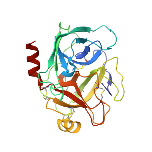
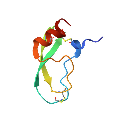
    Introducing fluorine into molecules has a wide range of effects on their physicochemical properties, often desirable but in most cases unpredictable. The fluorine atom imparts the C-F bond with low polarizability and high polarity, and significantly affects the behavior of neighboring functional groups, in a covalent or noncovalent manner. Here, we report that fluorine, present in the form of a single fluoroalkyl amino acid side chain in the P1 position of the well-characterized serine-protease inhibitor BPTI, can fully restore inhibitor activity to a mutant that contains the corresponding hydrocarbon side chain at the same site. High resolution crystal structures were obtained for four BPTI variants in complex with bovine β-trypsin, revealing changes in the stoichiometry and dynamics of water molecules in the S1 subsite. These results demonstrate that the introduction of fluorine into a protein environment can result in "chemical complementation" that has a significantly favorable impact on protein-protein interactions.


  • Organizational Affiliation

    Department of Biology, Chemistry, and Pharmacy , Freie Universität Berlin , Institute of Chemistry and Biochemistry , Takustr. 3 , Berlin, 14195 , Germany . Email: beate.koksch@fu-berlin.de ; ; Tel: +49-30-83855344.


Macromolecules
Find similar proteins by:  (by identity cutoff)  |  3D Structure
Entity ID: 1
MoleculeChains Sequence LengthOrganismDetailsImage
Cationic trypsinA [auth E]223Bos taurusMutation(s): 0 
EC: 3.4.21.4
UniProt
Find proteins for P00760 (Bos taurus)
Explore P00760 
Go to UniProtKB:  P00760
Entity Groups  
Sequence Clusters30% Identity50% Identity70% Identity90% Identity95% Identity100% Identity
UniProt GroupP00760
Sequence Annotations
Expand
  • Reference Sequence
Find similar proteins by:  (by identity cutoff)  |  3D Structure
Entity ID: 2
MoleculeChains Sequence LengthOrganismDetailsImage
Pancreatic trypsin inhibitorB [auth I]58Bos taurusMutation(s): 0 
UniProt
Find proteins for P00974 (Bos taurus)
Explore P00974 
Go to UniProtKB:  P00974
Entity Groups  
Sequence Clusters30% Identity50% Identity70% Identity90% Identity95% Identity100% Identity
UniProt GroupP00974
Sequence Annotations
Expand
  • Reference Sequence
Small Molecules
Ligands 3 Unique
IDChains Name / Formula / InChI Key2D Diagram3D Interactions
SO4
Query on SO4

Download Ideal Coordinates CCD File 
C [auth E]
D [auth E]
E
F [auth E]
J [auth I]
C [auth E],
D [auth E],
E,
F [auth E],
J [auth I],
K [auth I],
L [auth I],
M [auth I],
N [auth I]
SULFATE ION
O4 S
QAOWNCQODCNURD-UHFFFAOYSA-L
GOL
Query on GOL

Download Ideal Coordinates CCD File 
G [auth E],
H [auth E]
GLYCEROL
C3 H8 O3
PEDCQBHIVMGVHV-UHFFFAOYSA-N
CA
Query on CA

Download Ideal Coordinates CCD File 
I [auth E]CALCIUM ION
Ca
BHPQYMZQTOCNFJ-UHFFFAOYSA-N
Experimental Data & Validation

Experimental Data

  • Method: X-RAY DIFFRACTION
  • Resolution: 1.25 Å
  • R-Value Free: 0.155 
  • R-Value Work: 0.138 
  • R-Value Observed: 0.139 
  • Space Group: I 2 2 2
Unit Cell:
Length ( Å )Angle ( ˚ )
a = 74.8α = 90
b = 81.49β = 90
c = 124.21γ = 90
Software Package:
Software NamePurpose
PHENIXrefinement
XDSdata reduction
XSCALEdata scaling
PHASERphasing

Structure Validation

View Full Validation Report


Entry History & Funding Information

Deposition Data


Funding OrganizationLocationGrant Number
German Research FoundationGermanyGraduate 25 School GRK 1582/1

Revision History  (Full details and data files)

  • Version 1.0: 2015-06-24
    Type: Initial release
  • Version 1.1: 2015-08-26
    Changes: Database references
  • Version 2.0: 2018-04-18
    Changes: Advisory, Atomic model, Author supporting evidence, Data collection, Database references, Derived calculations
  • Version 2.1: 2024-01-10
    Changes: Data collection, Database references, Derived calculations, Refinement description