4XZU

Crystal Structure of the Human Factor VIII C2 Domain in Complex with Murine 3E6 Inhibitory Antibody


Experimental Data Snapshot

  • Method: X-RAY DIFFRACTION
  • Resolution: 2.61 Å
  • R-Value Free: 0.267 
  • R-Value Work: 0.199 
  • R-Value Observed: 0.202 

wwPDB Validation   3D Report Full Report


This is version 1.3 of the entry. See complete history


Literature

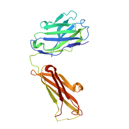
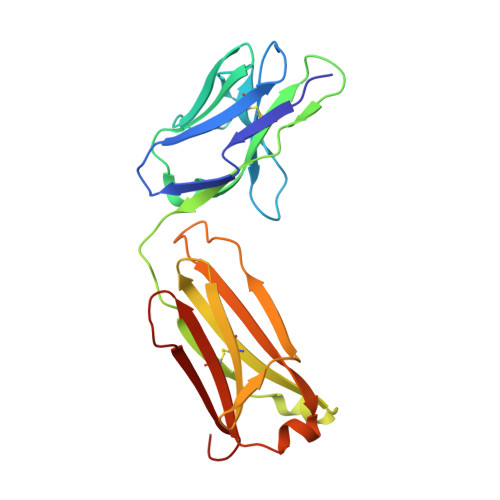
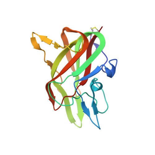
Structure of the Human Factor VIII C2 Domain in Complex with the 3E6 Inhibitory Antibody.

Wuerth, M.E.Cragerud, R.K.Clint Spiegel, P.

(2015) Sci Rep 5: 17216-17216

  • DOI: https://doi.org/10.1038/srep17216
  • Primary Citation of Related Structures:  
    4XZU

  • PubMed Abstract: 

    Blood coagulation factor VIII is a glycoprotein cofactor that is essential for the intrinsic pathway of the blood coagulation cascade. Inhibitory antibodies arise either spontaneously or in response to therapeutic infusion of functional factor VIII into hemophilia A patients, many of which are specific to the factor VIII C2 domain. The immune response is largely parsed into "classical" and "non-classical" inhibitory antibodies, which bind to opposing faces cooperatively. In this study, the 2.61 Å resolution structure of the C2 domain in complex with the antigen-binding fragment of the 3E6 classical inhibitory antibody is reported. The binding interface is largely conserved when aligned with the previously determined structure of the C2 domain in complex with two antibodies simultaneously. Further inspection of the B factors for the C2 domain in various X-ray crystal structures indicates that 3E6 antibody binding decreases the thermal motion behavior of surface loops in the C2 domain on the opposing face, thereby suggesting that cooperative antibody binding is a dynamic effect. Understanding the structural nature of the immune response to factor VIII following hemophilia A treatment will help lead to the development of better therapeutic reagents.


  • Organizational Affiliation

    Western Washington University, Department of Chemistry, 516 High Street, Bellingham, WA 98225-9150.


Macromolecules
Find similar proteins by:  (by identity cutoff)  |  3D Structure
Entity ID: 1
MoleculeChains Sequence LengthOrganismDetailsImage
3E6 antibody Fab heavy chainA,
D [auth E]
219Mus musculusMutation(s): 0 
Entity Groups  
Sequence Clusters30% Identity50% Identity70% Identity90% Identity95% Identity100% Identity
Sequence Annotations
Expand
  • Reference Sequence
Find similar proteins by:  (by identity cutoff)  |  3D Structure
Entity ID: 2
MoleculeChains Sequence LengthOrganismDetailsImage
3E6 antibody Fab light chainB,
E [auth F]
213Mus musculusMutation(s): 0 
Entity Groups  
Sequence Clusters30% Identity50% Identity70% Identity90% Identity95% Identity100% Identity
Sequence Annotations
Expand
  • Reference Sequence
Find similar proteins by:  (by identity cutoff)  |  3D Structure
Entity ID: 3
MoleculeChains Sequence LengthOrganismDetailsImage
Coagulation factor VIIIC [auth M],
F [auth G]
154Homo sapiensMutation(s): 0 
Gene Names: F8F8C
UniProt & NIH Common Fund Data Resources
Find proteins for P00451 (Homo sapiens)
Explore P00451 
Go to UniProtKB:  P00451
PHAROS:  P00451
GTEx:  ENSG00000185010 
Entity Groups  
Sequence Clusters30% Identity50% Identity70% Identity90% Identity95% Identity100% Identity
UniProt GroupP00451
Sequence Annotations
Expand
  • Reference Sequence
Experimental Data & Validation

Experimental Data

  • Method: X-RAY DIFFRACTION
  • Resolution: 2.61 Å
  • R-Value Free: 0.267 
  • R-Value Work: 0.199 
  • R-Value Observed: 0.202 
  • Space Group: P 2 21 21
Unit Cell:
Length ( Å )Angle ( ˚ )
a = 43.229α = 90
b = 148.492β = 90
c = 188.417γ = 90
Software Package:
Software NamePurpose
PHENIXrefinement
HKL-2000data reduction
PHASERphasing
PDB_EXTRACTdata extraction
HKL-2000data scaling
CrystalCleardata collection

Structure Validation

View Full Validation Report


Entry History & Funding Information

Deposition Data


Funding OrganizationLocationGrant Number
National Institutes of Health/National Heart, Lung, and Blood Institute (NIH/NHLBI)United StatesR15HL103518-01

Revision History  (Full details and data files)

  • Version 1.0: 2015-12-09
    Type: Initial release
  • Version 1.1: 2015-12-16
    Changes: Derived calculations
  • Version 1.2: 2017-09-13
    Changes: Author supporting evidence, Derived calculations
  • Version 1.3: 2019-12-04
    Changes: Author supporting evidence