4XYP

Crystal structure of a piscine viral fusion protein


Experimental Data Snapshot

  • Method: X-RAY DIFFRACTION
  • Resolution: 2.10 Å
  • R-Value Free: 0.251 
  • R-Value Work: 0.196 
  • R-Value Observed: 0.201 

wwPDB Validation   3D Report Full Report


Ligand Structure Quality Assessment 


This is version 1.5 of the entry. See complete history


Literature

Electrostatic Architecture of the Infectious Salmon Anemia Virus (ISAV) Core Fusion Protein Illustrates a Carboxyl-Carboxylate pH Sensor.

Cook, J.D.Soto-Montoya, H.Korpela, M.K.Lee, J.E.

(2015) J Biol Chem 290: 18495-18504

  • DOI: https://doi.org/10.1074/jbc.M115.644781
  • Primary Citation of Related Structures:  
    4XYP

  • PubMed Abstract: 

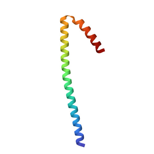
    Segment 5, ORF 1 of the infectious salmon anemia virus (ISAV) genome, encodes for the ISAV F protein, which is responsible for viral-host endosomal membrane fusion during a productive ISAV infection. The entry machinery of ISAV is composed of a complex of the ISAV F and ISAV hemagglutinin esterase (HE) proteins in an unknown stoichiometry prior to receptor engagement by ISAV HE. Following binding of the receptor to ISAV HE, dissociation of the ISAV F protein from HE, and subsequent endocytosis, the ISAV F protein resolves into a fusion-competent oligomeric state. Here, we present a 2.1 Å crystal structure of the fusion core of the ISAV F protein determined at low pH. This structure has allowed us to unambiguously demonstrate that the ISAV entry machinery exhibits typical class I viral fusion protein architecture. Furthermore, we have determined stabilizing factors that accommodate the pH-dependent mode of ISAV transmission, and our structure has allowed the identification of a central coil that is conserved across numerous and varied post-fusion viral glycoprotein structures. We then discuss a mechanistic model of ISAV fusion that parallels the paramyxoviral class I fusion strategy wherein attachment and fusion are relegated to separate proteins in a similar fashion to ISAV fusion.


  • Organizational Affiliation

    From the Department of Laboratory Medicine and Pathobiology, Faculty of Medicine, University of Toronto, Toronto, Ontario M5S 1A8, Canada.


Macromolecules
Find similar proteins by:  (by identity cutoff)  |  3D Structure
Entity ID: 1
MoleculeChains Sequence LengthOrganismDetailsImage
Fusion protein70Isavirus salarisMutation(s): 0 
UniProt
Find proteins for C6F339 (Isavirus salaris)
Explore C6F339 
Go to UniProtKB:  C6F339
Entity Groups  
Sequence Clusters30% Identity50% Identity70% Identity90% Identity95% Identity100% Identity
UniProt GroupC6F339
Sequence Annotations
Expand
  • Reference Sequence
Experimental Data & Validation

Experimental Data

  • Method: X-RAY DIFFRACTION
  • Resolution: 2.10 Å
  • R-Value Free: 0.251 
  • R-Value Work: 0.196 
  • R-Value Observed: 0.201 
  • Space Group: H 3 2
Unit Cell:
Length ( Å )Angle ( ˚ )
a = 42.874α = 90
b = 42.874β = 90
c = 232.245γ = 120
Software Package:
Software NamePurpose
PHASERphasing
PHENIXrefinement
PDB_EXTRACTdata extraction
Aimlessdata scaling
Cootmodel building

Structure Validation

View Full Validation Report


Ligand Structure Quality Assessment 


Entry History & Funding Information

Deposition Data


Funding OrganizationLocationGrant Number
Natural Sciences and Engineering Research Council (NSERC, Canada)CanadaRGPIN435607-13

Revision History  (Full details and data files)

  • Version 1.0: 2015-06-24
    Type: Initial release
  • Version 1.1: 2015-07-01
    Changes: Database references
  • Version 1.2: 2015-08-05
    Changes: Database references, Other
  • Version 1.3: 2017-09-06
    Changes: Author supporting evidence, Derived calculations
  • Version 1.4: 2020-01-08
    Changes: Author supporting evidence
  • Version 1.5: 2024-02-28
    Changes: Data collection, Database references