4XUI

Crystal structure analysis of cruzain bound to the no-covalent analog of WRR-483 (WRR-669)


Experimental Data Snapshot

  • Method: X-RAY DIFFRACTION
  • Resolution: 2.51 Å
  • R-Value Free: 0.189 
  • R-Value Work: 0.160 
  • R-Value Observed: 0.163 

wwPDB Validation   3D Report Full Report


Ligand Structure Quality Assessment 


This is version 1.2 of the entry. See complete history


Literature

Synthesis and Evaluation of Oxyguanidine Analogues of the Cysteine Protease Inhibitor WRR-483 against Cruzain.

Jones, B.D.Tochowicz, A.Tang, Y.Cameron, M.D.McCall, L.I.Hirata, K.Siqueira-Neto, J.L.Reed, S.L.McKerrow, J.H.Roush, W.R.

(2016) ACS Med Chem Lett 7: 77-82

  • DOI: https://doi.org/10.1021/acsmedchemlett.5b00336
  • Primary Citation of Related Structures:  
    4PI3, 4XUI

  • PubMed Abstract: 

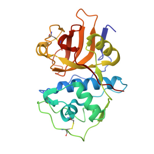
    A series of oxyguanidine analogues of the cysteine protease inhibitor WRR-483 were synthesized and evaluated against cruzain, the major cysteine protease of the protozoan parasite Trypanosoma cruzi. Kinetic analyses of these analogues indicated that they have comparable potency to previously prepared vinyl sulfone cruzain inhibitors. Co-crystal structures of the oxyguanidine analogues WRR-666 (4) and WRR-669 (7) bound to cruzain demonstrated different binding interactions with the cysteine protease, depending on the aryl moiety of the P1' inhibitor subunit. Specifically, these data demonstrate that WRR-669 is bound noncovalently in the crystal structure. This represents a rare example of noncovalent inhibition of a cysteine protease by a vinyl sulfone inhibitor.


  • Organizational Affiliation

    Department of Chemistry, The Scripps Research Institute , 130 Scripps Way, Jupiter, Florida 33458, United States.


Macromolecules
Find similar proteins by:  (by identity cutoff)  |  3D Structure
Entity ID: 1
MoleculeChains Sequence LengthOrganismDetailsImage
Cruzipain
A, B, C
216Trypanosoma cruziMutation(s): 0 
EC: 3.4.22.51
UniProt
Find proteins for P25779 (Trypanosoma cruzi)
Explore P25779 
Go to UniProtKB:  P25779
Entity Groups  
Sequence Clusters30% Identity50% Identity70% Identity90% Identity95% Identity100% Identity
UniProt GroupP25779
Sequence Annotations
Expand
  • Reference Sequence
Experimental Data & Validation

Experimental Data

  • Method: X-RAY DIFFRACTION
  • Resolution: 2.51 Å
  • R-Value Free: 0.189 
  • R-Value Work: 0.160 
  • R-Value Observed: 0.163 
  • Space Group: P 32
Unit Cell:
Length ( Å )Angle ( ˚ )
a = 101.2α = 90
b = 101.2β = 90
c = 64.9γ = 120
Software Package:
Software NamePurpose
PHENIXrefinement
XDSdata reduction
XDSdata scaling
PHASERphasing

Structure Validation

View Full Validation Report


Ligand Structure Quality Assessment 


Entry History 

Deposition Data

Revision History  (Full details and data files)

  • Version 1.0: 2015-03-11
    Type: Initial release
  • Version 1.1: 2016-02-10
    Changes: Database references
  • Version 1.2: 2023-09-27
    Changes: Data collection, Database references, Derived calculations, Refinement description