4XP7

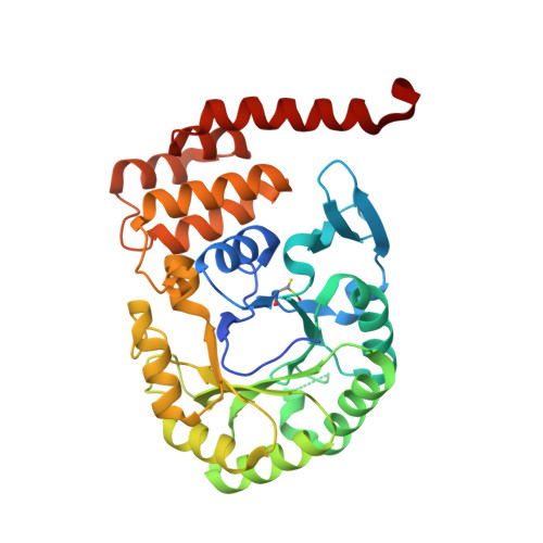
Crystal structure of Human tRNA dihydrouridine synthase 2


Experimental Data Snapshot

  • Method: X-RAY DIFFRACTION
  • Resolution: 1.90 Å
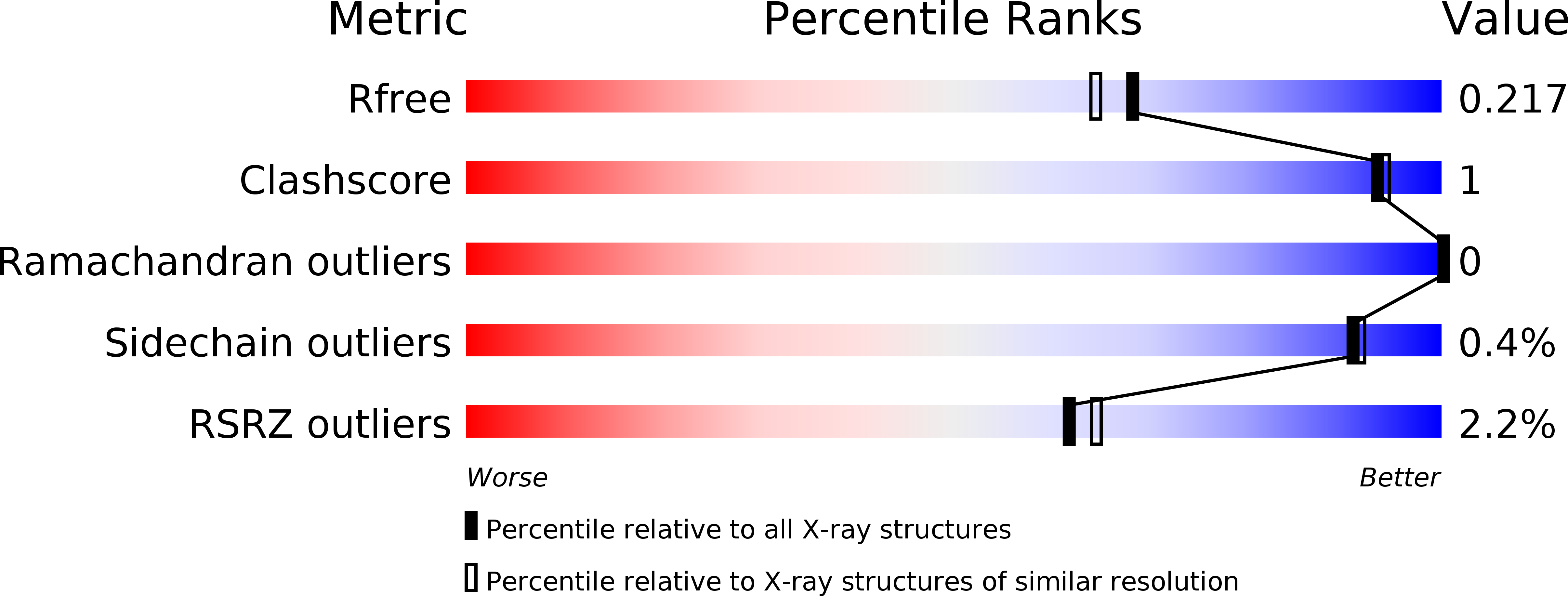
  • R-Value Free: 0.206 
  • R-Value Work: 0.168 
  • R-Value Observed: 0.170 

wwPDB Validation   3D Report Full Report


Ligand Structure Quality Assessment 


This is version 1.1 of the entry. See complete history


Literature

From bacterial to human dihydrouridine synthase: automated structure determination.

Whelan, F.Jenkins, H.T.Griffiths, S.C.Byrne, R.T.Dodson, E.J.Antson, A.A.

(2015) Acta Crystallogr D Biol Crystallogr 71: 1564-1571

  • DOI: https://doi.org/10.1107/S1399004715009220
  • Primary Citation of Related Structures:  
    4XP7

  • PubMed Abstract: 

    The reduction of uridine to dihydrouridine at specific positions in tRNA is catalysed by dihydrouridine synthase (Dus) enzymes. Increased expression of human dihydrouridine synthase 2 (hDus2) has been linked to pulmonary carcinogenesis, while its knockdown decreased cancer cell line viability, suggesting that it may serve as a valuable target for therapeutic intervention. Here, the X-ray crystal structure of a construct of hDus2 encompassing the catalytic and tRNA-recognition domains (residues 1-340) determined at 1.9 Å resolution is presented. It is shown that the structure can be determined automatically by phenix.mr_rosetta starting from a bacterial Dus enzyme with only 18% sequence identity and a significantly divergent structure. The overall fold of the human Dus2 is similar to that of bacterial enzymes, but has a larger recognition domain and a unique three-stranded antiparallel β-sheet insertion into the catalytic domain that packs next to the recognition domain, contributing to domain-domain interactions. The structure may inform the development of novel therapeutic approaches in the fight against lung cancer.


  • Organizational Affiliation

    Department of Biology, The University of York, Heslington, York YO10 5DD, England.


Macromolecules
Find similar proteins by:  (by identity cutoff)  |  3D Structure
Entity ID: 1
MoleculeChains Sequence LengthOrganismDetailsImage
tRNA-dihydrouridine(20) synthase [NAD(P)+]-like346Homo sapiensMutation(s): 0 
Gene Names: DUS2DUS2L
EC: 1.3.1
UniProt & NIH Common Fund Data Resources
Find proteins for Q9NX74 (Homo sapiens)
Explore Q9NX74 
Go to UniProtKB:  Q9NX74
PHAROS:  Q9NX74
GTEx:  ENSG00000167264 
Entity Groups  
Sequence Clusters30% Identity50% Identity70% Identity90% Identity95% Identity100% Identity
UniProt GroupQ9NX74
Sequence Annotations
Expand
  • Reference Sequence
Small Molecules
Ligands 1 Unique
IDChains Name / Formula / InChI Key2D Diagram3D Interactions
FNR
Query on FNR

Download Ideal Coordinates CCD File 
B [auth A]1-DEOXY-1-(7,8-DIMETHYL-2,4-DIOXO-3,4-DIHYDRO-2H-BENZO[G]PTERIDIN-1-ID-10(5H)-YL)-5-O-PHOSPHONATO-D-RIBITOL
C17 H23 N4 O9 P
YTNIXZGTHTVJBW-SCRDCRAPSA-N
Modified Residues  1 Unique
IDChains TypeFormula2D DiagramParent
CSO
Query on CSO
A
L-PEPTIDE LINKINGC3 H7 N O3 SCYS
Experimental Data & Validation

Experimental Data

  • Method: X-RAY DIFFRACTION
  • Resolution: 1.90 Å
  • R-Value Free: 0.206 
  • R-Value Work: 0.168 
  • R-Value Observed: 0.170 
  • Space Group: P 1 21 1
Unit Cell:
Length ( Å )Angle ( ˚ )
a = 46.252α = 90
b = 49.562β = 95.71
c = 69.612γ = 90
Software Package:
Software NamePurpose
REFMACrefinement
SCALAdata scaling
MOLREPphasing
PDB_EXTRACTdata extraction
XDSdata reduction

Structure Validation

View Full Validation Report


Ligand Structure Quality Assessment 


Entry History & Funding Information

Deposition Data


Funding OrganizationLocationGrant Number
Wellcome TrustUnited Kingdom081916
Wellcome TrustUnited Kingdom098230

Revision History  (Full details and data files)

  • Version 1.0: 2015-01-28
    Type: Initial release
  • Version 1.1: 2015-07-15
    Changes: Database references