4XO9

Crystal structure of a FimH*DsG complex from E.coli K12 in space group C2


Experimental Data Snapshot

  • Method: X-RAY DIFFRACTION
  • Resolution: 1.14 Å
  • R-Value Free: 0.143 
  • R-Value Work: 0.125 
  • R-Value Observed: 0.125 

wwPDB Validation   3D Report Full Report


This is version 1.2 of the entry. See complete history


Literature

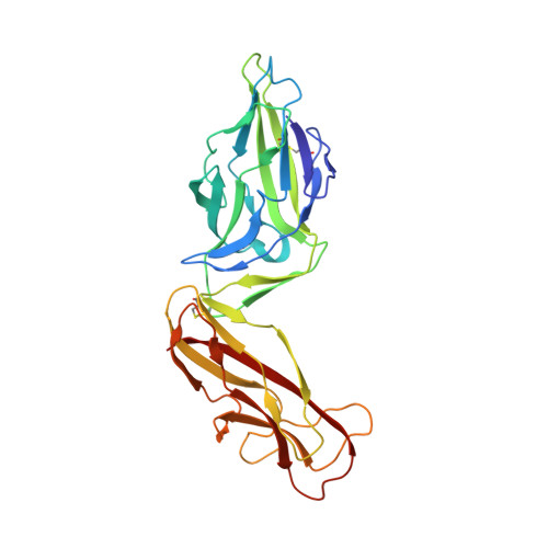
Catch-bond mechanism of the bacterial adhesin FimH.

Sauer, M.M.Jakob, R.P.Eras, J.Baday, S.Eris, D.Navarra, G.Berneche, S.Ernst, B.Maier, T.Glockshuber, R.

(2016) Nat Commun 7: 10738-10738

  • DOI: https://doi.org/10.1038/ncomms10738
  • Primary Citation of Related Structures:  
    4XO8, 4XO9, 4XOA, 4XOB, 4XOC, 4XOD, 4XOE

  • PubMed Abstract: 

    Ligand-receptor interactions that are reinforced by mechanical stress, so-called catch-bonds, play a major role in cell-cell adhesion. They critically contribute to widespread urinary tract infections by pathogenic Escherichia coli strains. These pathogens attach to host epithelia via the adhesin FimH, a two-domain protein at the tip of type I pili recognizing terminal mannoses on epithelial glycoproteins. Here we establish peptide-complemented FimH as a model system for fimbrial FimH function. We reveal a three-state mechanism of FimH catch-bond formation based on crystal structures of all states, kinetic analysis of ligand interaction and molecular dynamics simulations. In the absence of tensile force, the FimH pilin domain allosterically accelerates spontaneous ligand dissociation from the FimH lectin domain by 100,000-fold, resulting in weak affinity. Separation of the FimH domains under stress abolishes allosteric interplay and increases the affinity of the lectin domain. Cell tracking demonstrates that rapid ligand dissociation from FimH supports motility of piliated E. coli on mannosylated surfaces in the absence of shear force.


  • Organizational Affiliation

    Institute of Molecular Biology and Biophysics, Department of Biology, ETH, Zurich, Otto-Stern-Weg 5, 8093 Zurich, Switzerland.


Macromolecules
Find similar proteins by:  (by identity cutoff)  |  3D Structure
Entity ID: 1
MoleculeChains Sequence LengthOrganismDetailsImage
Protein FimH279Escherichia coli K-12Mutation(s): 0 
Gene Names: fimHb4320JW4283
UniProt
Find proteins for P08191 (Escherichia coli (strain K12))
Explore P08191 
Go to UniProtKB:  P08191
Entity Groups  
Sequence Clusters30% Identity50% Identity70% Identity90% Identity95% Identity100% Identity
UniProt GroupP08191
Sequence Annotations
Expand
  • Reference Sequence

Find similar proteins by:  Sequence   |   3D Structure  

Entity ID: 2
MoleculeChains Sequence LengthOrganismDetailsImage
Minor component of type 1 fimbriae14Escherichia coli K-12Mutation(s): 0 
Gene Names: fimGBWG_4017
UniProt
Find proteins for P08190 (Escherichia coli (strain K12))
Explore P08190 
Go to UniProtKB:  P08190
Entity Groups  
Sequence Clusters30% Identity50% Identity70% Identity90% Identity95% Identity100% Identity
UniProt GroupP08190
Sequence Annotations
Expand
  • Reference Sequence
Experimental Data & Validation

Experimental Data

  • Method: X-RAY DIFFRACTION
  • Resolution: 1.14 Å
  • R-Value Free: 0.143 
  • R-Value Work: 0.125 
  • R-Value Observed: 0.125 
  • Space Group: C 1 2 1
Unit Cell:
Length ( Å )Angle ( ˚ )
a = 99.49α = 90
b = 35.62β = 105.89
c = 72.89γ = 90
Software Package:
Software NamePurpose
PHENIXrefinement
XDSdata reduction
XSCALEdata scaling
PHASERphasing

Structure Validation

View Full Validation Report


Entry History 

Deposition Data

Revision History  (Full details and data files)

  • Version 1.0: 2016-01-27
    Type: Initial release
  • Version 1.1: 2016-03-16
    Changes: Database references
  • Version 1.2: 2024-01-10
    Changes: Data collection, Database references, Derived calculations, Refinement description