4XF7

myo-inositol 3-kinase bound with its substrates (AMPPCP and myo-inositol)


Experimental Data Snapshot

  • Method: X-RAY DIFFRACTION
  • Resolution: 1.93 Å
  • R-Value Free: 0.255 
  • R-Value Work: 0.216 
  • R-Value Observed: 0.218 

wwPDB Validation   3D Report Full Report


Ligand Structure Quality Assessment 


This is version 1.4 of the entry. See complete history


Literature

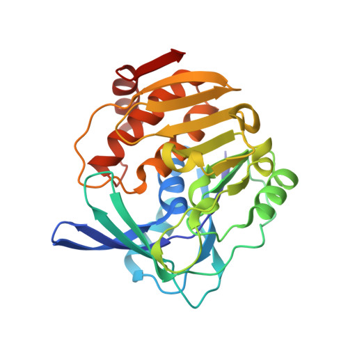
Crystal Structure and Product Analysis of an Archaeal myo-Inositol Kinase Reveal Substrate Recognition Mode and 3-OH Phosphorylation

Nagata, R.Fujihashi, M.Sato, T.Atomi, H.Miki, K.

(2015) Biochemistry 54: 3494-3503

  • DOI: https://doi.org/10.1021/acs.biochem.5b00296
  • Primary Citation of Related Structures:  
    4XF6, 4XF7

  • PubMed Abstract: 

    The TK2285 protein from Thermococcus kodakarensis was recently characterized as an enzyme catalyzing the phosphorylation of myo-inositol. Only two myo-inositol kinases have been identified so far, the TK2285 protein and Lpa3 from Zea mays, both of which belong to the ribokinase family. In either case, which of the six hydroxyl groups of myo-inositol is phosphorylated is still unknown. In addition, little is known about the myo-inositol binding mechanism of these enzymes. In this work, we determined two crystal structures: those of the TK2285 protein complexed with the substrates (ATP analogue and myo-inositol) or the reaction products formed by the enzyme. Analysis of the ternary substrates-complex structure and site-directed mutagenesis showed that five residues were involved in the interaction with myo-inositol. Structural comparison with other ribokinase family enzymes indicated that two of the five residues, Q136 and R140, are characteristic of myo-inositol kinase. The crystal structure of the ternary products-complex, which was prepared by incubating the TK2285 protein with myo-inositol and ATP, holds 1d-myo-inositol 3-phosphate (Ins(3)P) in the active site. NMR and HPLC analyses with a chiral column also indicated that the TK2285 reaction product was Ins(3)P. The results obtained here showed that the TK2285 protein specifically catalyzes the phosphorylation of the 3-OH of myo-inositol. We thus designated TK2285 as myo-inositol 3-kinase (MI3K). The precise identification of the reaction product should provide a sound basis to further explore inositol metabolism in Archaea.


  • Organizational Affiliation

    †Department of Chemistry, Graduate School of Science, Kyoto University, Sakyo-ku, Kyoto 606-8502, Japan.


Macromolecules
Find similar proteins by:  (by identity cutoff)  |  3D Structure
Entity ID: 1
MoleculeChains Sequence LengthOrganismDetailsImage
Carbohydrate/pyrimidine kinase, PfkB family
A, B
273Thermococcus kodakarensis KOD1Mutation(s): 0 
Gene Names: TK2285
EC: 2.7.1.64
UniProt
Find proteins for Q5JDA3 (Thermococcus kodakarensis (strain ATCC BAA-918 / JCM 12380 / KOD1))
Explore Q5JDA3 
Go to UniProtKB:  Q5JDA3
Entity Groups  
Sequence Clusters30% Identity50% Identity70% Identity90% Identity95% Identity100% Identity
UniProt GroupQ5JDA3
Sequence Annotations
Expand
  • Reference Sequence
Small Molecules
Ligands 4 Unique
IDChains Name / Formula / InChI Key2D Diagram3D Interactions
ACP
Query on ACP

Download Ideal Coordinates CCD File 
D [auth A],
S [auth B]
PHOSPHOMETHYLPHOSPHONIC ACID ADENYLATE ESTER
C11 H18 N5 O12 P3
UFZTZBNSLXELAL-IOSLPCCCSA-N
INS
Query on INS

Download Ideal Coordinates CCD File 
C [auth A],
R [auth B]
1,2,3,4,5,6-HEXAHYDROXY-CYCLOHEXANE
C6 H12 O6
CDAISMWEOUEBRE-GPIVLXJGSA-N
IOD
Query on IOD

Download Ideal Coordinates CCD File 
AA [auth B]
BA [auth B]
CA [auth B]
DA [auth B]
F [auth A]
AA [auth B],
BA [auth B],
CA [auth B],
DA [auth B],
F [auth A],
G [auth A],
H [auth A],
I [auth A],
J [auth A],
K [auth A],
L [auth A],
M [auth A],
N [auth A],
O [auth A],
P [auth A],
Q [auth A],
U [auth B],
V [auth B],
W [auth B],
X [auth B],
Y [auth B],
Z [auth B]
IODIDE ION
I
XMBWDFGMSWQBCA-UHFFFAOYSA-M
MG
Query on MG

Download Ideal Coordinates CCD File 
E [auth A],
T [auth B]
MAGNESIUM ION
Mg
JLVVSXFLKOJNIY-UHFFFAOYSA-N
Experimental Data & Validation

Experimental Data

  • Method: X-RAY DIFFRACTION
  • Resolution: 1.93 Å
  • R-Value Free: 0.255 
  • R-Value Work: 0.216 
  • R-Value Observed: 0.218 
  • Space Group: P 21 21 21
Unit Cell:
Length ( Å )Angle ( ˚ )
a = 77.134α = 90
b = 81.176β = 90
c = 81.109γ = 90
Software Package:
Software NamePurpose
REFMACrefinement
PDB_EXTRACTdata extraction
HKL-2000data scaling
REFMACphasing

Structure Validation

View Full Validation Report


Ligand Structure Quality Assessment 


Entry History 

Deposition Data

Revision History  (Full details and data files)

  • Version 1.0: 2015-06-03
    Type: Initial release
  • Version 1.1: 2015-06-10
    Changes: Database references
  • Version 1.2: 2015-06-24
    Changes: Database references
  • Version 1.3: 2018-03-28
    Changes: Data collection, Database references, Derived calculations, Experimental preparation, Source and taxonomy
  • Version 1.4: 2023-11-08
    Changes: Data collection, Database references, Derived calculations, Refinement description