4XD7

Structure of thermophilic F1-ATPase inhibited by epsilon subunit


Experimental Data Snapshot

  • Method: X-RAY DIFFRACTION
  • Resolution: 3.90 Å
  • R-Value Free: 0.282 
  • R-Value Work: 0.250 
  • R-Value Observed: 0.251 

wwPDB Validation   3D Report Full Report


Ligand Structure Quality Assessment 


This is version 1.4 of the entry. See complete history


Literature

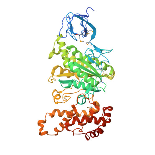
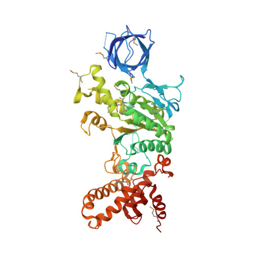
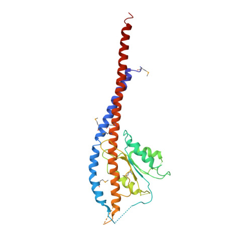
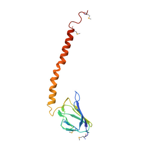
Structure of a thermophilic F1 -ATPase inhibited by an epsilon-subunit: deeper insight into the epsilon-inhibition mechanism.

Shirakihara, Y.Shiratori, A.Tanikawa, H.Nakasako, M.Yoshida, M.Suzuki, T.

(2015) FEBS J 282: 2895-2913

  • DOI: https://doi.org/10.1111/febs.13329
  • Primary Citation of Related Structures:  
    4XD7

  • PubMed Abstract: 

    F1-ATPase (F1) is the catalytic sector in F(o)F1-ATP synthase that is responsible for ATP production in living cells. In catalysis, its three catalytic β-subunits undergo nucleotide occupancy-dependent and concerted open-close conformational changes that are accompanied by rotation of the γ-subunit. Bacterial and chloroplast F1 are inhibited by their own ε-subunit. In the ε-inhibited Escherichia coli F1 structure, the ε-subunit stabilizes the overall conformation (half-closed, closed, open) of the β-subunits by inserting its C-terminal helix into the α3β3 cavity. The structure of ε-inhibited thermophilic F1 is similar to that of E. coli F1, showing a similar conformation of the ε-subunit, but the thermophilic ε-subunit stabilizes another unique overall conformation (open, closed, open) of the β-subunits. The ε-C-terminal helix 2 and hook are conserved between the two structures in interactions with target residues and in their positions. Rest of the ε-C-terminal domains are in quite different conformations and positions, and have different modes of interaction with targets. This region is thought to serve ε-inhibition differently. For inhibition, the ε-subunit contacts the second catches of some of the β- and α-subunits, the N- and C-terminal helices, and some of the Rossmann fold segments. Those contacts, as a whole, lead to positioning of those β- and α- second catches in ε-inhibition-specific positions, and prevent rotation of the γ-subunit. Some of the structural features are observed even in IF1 inhibition in mitochondrial F1.


  • Organizational Affiliation

    National Institute of Genetics, Mishima, Japan.


Macromolecules
Find similar proteins by:  (by identity cutoff)  |  3D Structure
Entity ID: 1
MoleculeChains Sequence LengthOrganismDetailsImage
ATP synthase subunit alpha
A, B, C
502Bacillus sp. PS3Mutation(s): 0 
Gene Names: atpA
EC: 3.6.3.14
UniProt
Find proteins for P09219 (Bacillus sp. (strain PS3))
Explore P09219 
Go to UniProtKB:  P09219
Entity Groups  
Sequence Clusters30% Identity50% Identity70% Identity90% Identity95% Identity100% Identity
UniProt GroupP09219
Sequence Annotations
Expand
  • Reference Sequence
Find similar proteins by:  (by identity cutoff)  |  3D Structure
Entity ID: 2
MoleculeChains Sequence LengthOrganismDetailsImage
ATP synthase subunit beta
D, E, F
483Bacillus sp. PS3Mutation(s): 0 
Gene Names: atpD
EC: 3.6.3.14
UniProt
Find proteins for P07677 (Bacillus sp. (strain PS3))
Explore P07677 
Go to UniProtKB:  P07677
Entity Groups  
Sequence Clusters30% Identity50% Identity70% Identity90% Identity95% Identity100% Identity
UniProt GroupP07677
Sequence Annotations
Expand
  • Reference Sequence
Find similar proteins by:  (by identity cutoff)  |  3D Structure
Entity ID: 3
MoleculeChains Sequence LengthOrganismDetailsImage
ATP synthase gamma chain285Bacillus sp. PS3Mutation(s): 0 
Gene Names: atpG
UniProt
Find proteins for P09222 (Bacillus sp. (strain PS3))
Explore P09222 
Go to UniProtKB:  P09222
Entity Groups  
Sequence Clusters30% Identity50% Identity70% Identity90% Identity95% Identity100% Identity
UniProt GroupP09222
Sequence Annotations
Expand
  • Reference Sequence
Find similar proteins by:  (by identity cutoff)  |  3D Structure
Entity ID: 4
MoleculeChains Sequence LengthOrganismDetailsImage
ATP synthase epsilon chain133Bacillus sp. PS3Mutation(s): 0 
Gene Names: atpC
UniProt
Find proteins for P07678 (Bacillus sp. (strain PS3))
Explore P07678 
Go to UniProtKB:  P07678
Entity Groups  
Sequence Clusters30% Identity50% Identity70% Identity90% Identity95% Identity100% Identity
UniProt GroupP07678
Sequence Annotations
Expand
  • Reference Sequence
Experimental Data & Validation

Experimental Data

  • Method: X-RAY DIFFRACTION
  • Resolution: 3.90 Å
  • R-Value Free: 0.282 
  • R-Value Work: 0.250 
  • R-Value Observed: 0.251 
  • Space Group: I 41 2 2
Unit Cell:
Length ( Å )Angle ( ˚ )
a = 233.395α = 90
b = 233.395β = 90
c = 303.964γ = 90
Software Package:
Software NamePurpose
PHENIXrefinement
MOSFLMdata reduction
SCALAdata scaling
AMoREphasing

Structure Validation

View Full Validation Report


Ligand Structure Quality Assessment 


Entry History 

Deposition Data

Revision History  (Full details and data files)

  • Version 1.0: 2015-08-26
    Type: Initial release
  • Version 1.1: 2015-10-07
    Changes: Source and taxonomy
  • Version 1.2: 2017-04-19
    Changes: Structure summary
  • Version 1.3: 2023-09-27
    Changes: Advisory, Data collection, Database references, Refinement description
  • Version 1.4: 2023-11-15
    Changes: Data collection