4X7T

Structure of Omalizumab Fab fragment, crystal form 2


Experimental Data Snapshot

  • Method: X-RAY DIFFRACTION
  • Resolution: 3.00 Å
  • R-Value Free: 0.236 
  • R-Value Work: 0.193 
  • R-Value Observed: 0.197 

wwPDB Validation   3D Report Full Report


This is version 1.4 of the entry. See complete history


Literature

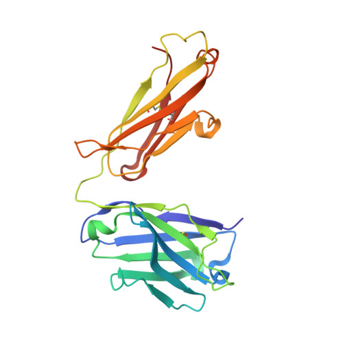
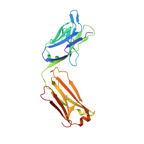
Structure of the omalizumab Fab

Jensen, R.K.Plum, M.Tjerrild, L.Jakob, T.Spillner, E.Andersen, G.R.

(2015) Acta Crystallogr F Struct Biol Commun 71: 419-426

  • DOI: https://doi.org/10.1107/S2053230X15004100
  • Primary Citation of Related Structures:  
    4X7S, 4X7T

  • PubMed Abstract: 

    Omalizumab is a humanized anti-IgE antibody that inhibits the binding of IgE to its receptors on mast cells and basophils, thus blocking the IgE-mediated release of inflammatory mediators from these cells. Omalizumab binds to the Fc domains of IgE in proximity to the binding site of the high-affinity IgE receptor FcℇRI, but the epitope and the mechanisms and conformations governing the recognition remain unknown. In order to elucidate the molecular mechanism of its anti-IgE activity, the aim was to analyse the interaction of omalizumab with human IgE. Therefore, IgE Fc Cℇ2-4 was recombinantly produced in mammalian HEK-293 cells. Functionality of the IgE Fc was proven by ELISA and mediator-release assays. Omalizumab IgG was cleaved with papain and the resulting Fab was purified by ion-exchange chromatography. The complex of IgE Fc with omalizumab was prepared by size-exclusion chromatography. However, crystals containing the complex were not obtained, suggesting that the process of crystallization favoured the dissociation of the two proteins. Instead, two structures of the omalizumab Fab with maximum resolutions of 1.9 and 3.0 Å were obtained. The structures reveal the arrangement of the CDRs and the position of omalizumab residues known from prior functional studies to be involved in IgE binding. Thus, the structure of omalizumab provides the structural basis for understanding the function of omalizumab, allows optimization of the procedure for complex crystallization and poses questions about the conformational requirements for anti-IgE activity.


  • Organizational Affiliation

    Department of Molecular Biology and Genetics, Aarhus University, Gustav Wiedsvej 10C, 8000 Aarhus, Denmark.


Macromolecules
Find similar proteins by:  (by identity cutoff)  |  3D Structure
Entity ID: 1
MoleculeChains Sequence LengthOrganismDetailsImage
Omalizumab-Fab Heavy chainA [auth H],
C [auth A]
222Homo sapiensMutation(s): 0 
Entity Groups  
Sequence Clusters30% Identity50% Identity70% Identity90% Identity95% Identity100% Identity
Sequence Annotations
Expand
  • Reference Sequence
Find similar proteins by:  (by identity cutoff)  |  3D Structure
Entity ID: 2
MoleculeChains Sequence LengthOrganismDetailsImage
Omalizumab-Fab Light chainB [auth L],
D [auth B]
218Homo sapiensMutation(s): 0 
Entity Groups  
Sequence Clusters30% Identity50% Identity70% Identity90% Identity95% Identity100% Identity
Sequence Annotations
Expand
  • Reference Sequence
Experimental Data & Validation

Experimental Data

  • Method: X-RAY DIFFRACTION
  • Resolution: 3.00 Å
  • R-Value Free: 0.236 
  • R-Value Work: 0.193 
  • R-Value Observed: 0.197 
  • Space Group: P 1 21 1
Unit Cell:
Length ( Å )Angle ( ˚ )
a = 85.47α = 90
b = 73.49β = 116.46
c = 87.04γ = 90
Software Package:
Software NamePurpose
PHENIXrefinement
XDSdata reduction
XSCALEdata scaling
PHASERphasing

Structure Validation

View Full Validation Report


Entry History & Funding Information

Deposition Data


Funding OrganizationLocationGrant Number
Danish Natural Science Research CouncilDenmark--
DanScattDenmark--
Novo Nordisk Foundation, Hallas-Mooller FellowshipDenmark--

Revision History  (Full details and data files)

  • Version 1.0: 2015-03-25
    Type: Initial release
  • Version 1.1: 2015-04-08
    Changes: Database references
  • Version 1.2: 2015-04-22
    Changes: Database references
  • Version 1.3: 2018-03-07
    Changes: Data collection
  • Version 1.4: 2024-01-10
    Changes: Author supporting evidence, Data collection, Database references, Refinement description