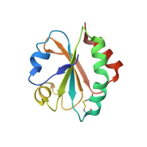
4X43

Structure of proline-free E. coli Thioredoxin


Experimental Data Snapshot

  • Method: X-RAY DIFFRACTION
  • Resolution: 1.65 Å
  • R-Value Free: 0.218 
  • R-Value Work: 0.176 
  • R-Value Observed: 0.177 

wwPDB Validation   3D Report Full Report


This is version 2.2 of the entry. See complete history


Literature

Acceleration of protein folding by four orders of magnitude through a single amino acid substitution.

Roderer, D.J.Scharer, M.A.Rubini, M.Glockshuber, R.

(2015) Sci Rep 5: 11840-11840

  • DOI: https://doi.org/10.1038/srep11840
  • Primary Citation of Related Structures:  
    4X43

  • PubMed Abstract: 

    Cis prolyl peptide bonds are conserved structural elements in numerous protein families, although their formation is energetically unfavorable, intrinsically slow and often rate-limiting for folding. Here we investigate the reasons underlying the conservation of the cis proline that is diagnostic for the fold of thioredoxin-like thiol-disulfide oxidoreductases. We show that replacement of the conserved cis proline in thioredoxin by alanine can accelerate spontaneous folding to the native, thermodynamically most stable state by more than four orders of magnitude. However, the resulting trans alanine bond leads to small structural rearrangements around the active site that impair the function of thioredoxin as catalyst of electron transfer reactions by more than 100-fold. Our data provide evidence for the absence of a strong evolutionary pressure to achieve intrinsically fast folding rates, which is most likely a consequence of proline isomerases and molecular chaperones that guarantee high in vivo folding rates and yields.


  • Organizational Affiliation

    ETH Zurich, Institute of Molecular Biology and Biophysics, Otto-Stern-Weg 5, CH-8093 Zurich, Switzerland.


Macromolecules
Find similar proteins by:  (by identity cutoff)  |  3D Structure
Entity ID: 1
MoleculeChains Sequence LengthOrganismDetailsImage
Thioredoxin-1
A, B, C
108Escherichia coli K-12Mutation(s): 5 
Gene Names: trxAfipAtsnCb3781JW5856
UniProt
Find proteins for P0AA25 (Escherichia coli (strain K12))
Explore P0AA25 
Go to UniProtKB:  P0AA25
Entity Groups  
Sequence Clusters30% Identity50% Identity70% Identity90% Identity95% Identity100% Identity
UniProt GroupP0AA25
Sequence Annotations
Expand
  • Reference Sequence
Experimental Data & Validation

Experimental Data

  • Method: X-RAY DIFFRACTION
  • Resolution: 1.65 Å
  • R-Value Free: 0.218 
  • R-Value Work: 0.176 
  • R-Value Observed: 0.177 
  • Space Group: P 1 21 1
Unit Cell:
Length ( Å )Angle ( ˚ )
a = 34.67α = 90
b = 48.57β = 101.01
c = 89.94γ = 90
Software Package:
Software NamePurpose
PHENIXrefinement
XDSdata reduction
XSCALEdata scaling
Cootmodel building
PDB_EXTRACTdata extraction
PHASERphasing

Structure Validation

View Full Validation Report


Entry History & Funding Information

Deposition Data


Funding OrganizationLocationGrant Number
ETH ZurichSwitzerland--
Swiss National Science FoundationSwitzerland--

Revision History  (Full details and data files)

  • Version 1.0: 2015-06-24
    Type: Initial release
  • Version 1.1: 2015-07-08
    Changes: Database references
  • Version 2.0: 2017-09-06
    Changes: Advisory, Atomic model, Author supporting evidence, Refinement description
  • Version 2.1: 2017-10-11
    Changes: Data collection
  • Version 2.2: 2024-01-10
    Changes: Data collection, Database references, Refinement description