4X2S

Crystal structure of 276S/M395R-GltPh in inward-facing conformation


Experimental Data Snapshot

  • Method: X-RAY DIFFRACTION
  • Resolution: 4.21 Å
  • R-Value Free: 0.314 
  • R-Value Work: 0.278 
  • R-Value Observed: 0.280 

wwPDB Validation   3D Report Full Report


This is version 1.4 of the entry. See complete history


Literature

Transport domain unlocking sets the uptake rate of an aspartate transporter.

Akyuz, N.Georgieva, E.R.Zhou, Z.Stolzenberg, S.Cuendet, M.A.Khelashvili, G.Altman, R.B.Terry, D.S.Freed, J.H.Weinstein, H.Boudker, O.Blanchard, S.C.

(2015) Nature 518: 68-73

  • DOI: https://doi.org/10.1038/nature14158
  • Primary Citation of Related Structures:  
    4X2S

  • PubMed Abstract: 

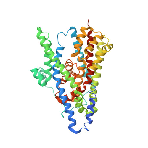
    Glutamate transporters terminate neurotransmission by clearing synaptically released glutamate from the extracellular space, allowing repeated rounds of signalling and preventing glutamate-mediated excitotoxicity. Crystallographic studies of a glutamate transporter homologue from the archaeon Pyrococcus horikoshii, GltPh, showed that distinct transport domains translocate substrates into the cytoplasm by moving across the membrane within a central trimerization scaffold. Here we report direct observations of these 'elevator-like' transport domain motions in the context of reconstituted proteoliposomes and physiological ion gradients using single-molecule fluorescence resonance energy transfer (smFRET) imaging. We show that GltPh bearing two mutations introduced to impart characteristics of the human transporter exhibits markedly increased transport domain dynamics, which parallels an increased rate of substrate transport, thereby establishing a direct temporal relationship between transport domain motion and substrate uptake. Crystallographic and computational investigations corroborated these findings by revealing that the 'humanizing' mutations favour structurally 'unlocked' intermediate states in the transport cycle exhibiting increased solvent occupancy at the interface between the transport domain and the trimeric scaffold.


  • Organizational Affiliation

    Department of Physiology and Biophysics, Weill Cornell Medical College, Cornell University, 1300 York Avenue, New York, New York 10065, USA.


Macromolecules
Find similar proteins by:  (by identity cutoff)  |  3D Structure
Entity ID: 1
MoleculeChains Sequence LengthOrganismDetailsImage
425aa long hypothetical proton glutamate symport protein
A, B, C
422Pyrococcus horikoshii OT3Mutation(s): 11 
Gene Names: PH1295
Membrane Entity: Yes 
UniProt
Find proteins for O59010 (Pyrococcus horikoshii (strain ATCC 700860 / DSM 12428 / JCM 9974 / NBRC 100139 / OT-3))
Explore O59010 
Go to UniProtKB:  O59010
Entity Groups  
Sequence Clusters30% Identity50% Identity70% Identity90% Identity95% Identity100% Identity
UniProt GroupO59010
Sequence Annotations
Expand
  • Reference Sequence
Experimental Data & Validation

Experimental Data

  • Method: X-RAY DIFFRACTION
  • Resolution: 4.21 Å
  • R-Value Free: 0.314 
  • R-Value Work: 0.278 
  • R-Value Observed: 0.280 
  • Space Group: P 65 2 2
Unit Cell:
Length ( Å )Angle ( ˚ )
a = 121.651α = 90
b = 121.651β = 90
c = 578.699γ = 120
Software Package:
Software NamePurpose
REFMACrefinement

Structure Validation

View Full Validation Report


Entry History & Funding Information

Deposition Data


Funding OrganizationLocationGrant Number
National Institutes of Health/National Institute of General Medical Sciences (NIH/NIGMS)United States5U54GM087519

Revision History  (Full details and data files)

  • Version 1.0: 2015-02-04
    Type: Initial release
  • Version 1.1: 2017-09-13
    Changes: Author supporting evidence, Derived calculations, Source and taxonomy
  • Version 1.2: 2018-04-25
    Changes: Data collection, Database references, Source and taxonomy
  • Version 1.3: 2019-12-25
    Changes: Author supporting evidence
  • Version 1.4: 2023-09-27
    Changes: Data collection, Database references, Refinement description