4X2R

Crystal structure of PriA from Actinomyces urogenitalis


Experimental Data Snapshot

  • Method: X-RAY DIFFRACTION
  • Resolution: 1.05 Å
  • R-Value Free: 0.141 
  • R-Value Work: 0.123 
  • R-Value Observed: 0.123 

wwPDB Validation   3D Report Full Report


Ligand Structure Quality Assessment 


This is version 1.4 of the entry. See complete history


Literature

Evolution of substrate specificity in a retained enzyme driven by gene loss.

Juarez-Vazquez, A.L.Edirisinghe, J.N.Verduzco-Castro, E.A.Michalska, K.Wu, C.Noda-Garcia, L.Babnigg, G.Endres, M.Medina-Ruiz, S.Santoyo-Flores, J.Carrillo-Tripp, M.Ton-That, H.Joachimiak, A.Henry, C.S.Barona-Gomez, F.

(2017) Elife 6

  • DOI: https://doi.org/10.7554/eLife.22679
  • Primary Citation of Related Structures:  
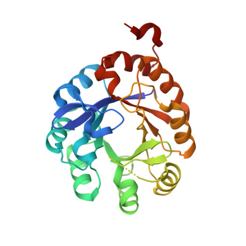
    4X2R

  • PubMed Abstract: 

    The connection between gene loss and the functional adaptation of retained proteins is still poorly understood. We apply phylogenomics and metabolic modeling to detect bacterial species that are evolving by gene loss, with the finding that Actinomycetaceae genomes from human cavities are undergoing sizable reductions, including loss of L-histidine and L-tryptophan biosynthesis. We observe that the dual-substrate phosphoribosyl isomerase A or priA gene, at which these pathways converge, appears to coevolve with the occurrence of trp and his genes. Characterization of a dozen PriA homologs shows that these enzymes adapt from bifunctionality in the largest genomes, to a monofunctional, yet not necessarily specialized, inefficient form in genomes undergoing reduction. These functional changes are accomplished via mutations, which result from relaxation of purifying selection, in residues structurally mapped after sequence and X-ray structural analyses. Our results show how gene loss can drive the evolution of substrate specificity from retained enzymes.


  • Organizational Affiliation

    Evolution of Metabolic Diversity Laboratory, Unidad de Genómica Avanzada (Langebio), Cinvestav-IPN, Irapuato, Mexico.


Macromolecules
Find similar proteins by:  (by identity cutoff)  |  3D Structure
Entity ID: 1
MoleculeChains Sequence LengthOrganismDetailsImage
1-(5-phosphoribosyl)-5-[(5-phosphoribosylamino)methylideneamino] imidazole-4-carboxamide isomerase250Actinomyces urogenitalis DSM 15434Mutation(s): 0 
Gene Names: hisAHMPREF0058_1848
EC: 5.3.1.16
UniProt
Find proteins for C0W7K4 (Actinomyces urogenitalis DSM 15434)
Explore C0W7K4 
Go to UniProtKB:  C0W7K4
Entity Groups  
Sequence Clusters30% Identity50% Identity70% Identity90% Identity95% Identity100% Identity
UniProt GroupC0W7K4
Sequence Annotations
Expand
  • Reference Sequence
Experimental Data & Validation

Experimental Data

  • Method: X-RAY DIFFRACTION
  • Resolution: 1.05 Å
  • R-Value Free: 0.141 
  • R-Value Work: 0.123 
  • R-Value Observed: 0.123 
  • Space Group: P 41
  • Diffraction Data: https://doi.org/10.18430/M34X2R
Unit Cell:
Length ( Å )Angle ( ˚ )
a = 76.02α = 90
b = 76.02β = 90
c = 43.179γ = 90
Software Package:
Software NamePurpose
PHENIXrefinement
HKL-3000data reduction
HKL-3000data scaling
PHASERphasing

Structure Validation

View Full Validation Report


Ligand Structure Quality Assessment 


Entry History & Funding Information

Deposition Data


Funding OrganizationLocationGrant Number
National Institutes of Health/National Institute of General Medical Sciences (NIH/NIGMS)United StatesGM094585

Revision History  (Full details and data files)

  • Version 1.0: 2014-12-24
    Type: Initial release
  • Version 1.1: 2017-09-20
    Changes: Advisory, Author supporting evidence, Derived calculations, Source and taxonomy, Structure summary
  • Version 1.2: 2017-11-08
    Changes: Database references
  • Version 1.3: 2019-12-25
    Changes: Author supporting evidence
  • Version 1.4: 2023-09-27
    Changes: Data collection, Database references, Refinement description