4WNT

Human Cytochrome P450 2D6 Ajmalicine Complex


Experimental Data Snapshot

  • Method: X-RAY DIFFRACTION
  • Resolution: 2.60 Å
  • R-Value Free: 0.237 
  • R-Value Work: 0.192 
  • R-Value Observed: 0.194 

wwPDB Validation   3D Report Full Report


Ligand Structure Quality Assessment 


This is version 1.6 of the entry. See complete history


Literature

Contributions of Ionic Interactions and Protein Dynamics to Cytochrome P450 2D6 (CYP2D6) Substrate and Inhibitor Binding.

Wang, A.Stout, C.D.Zhang, Q.Johnson, E.F.

(2015) J Biol Chem 290: 5092-5104

  • DOI: https://doi.org/10.1074/jbc.M114.627661
  • Primary Citation of Related Structures:  
    3TBG, 3TDA, 4WNT, 4WNU, 4WNV, 4WNW

  • PubMed Abstract: 

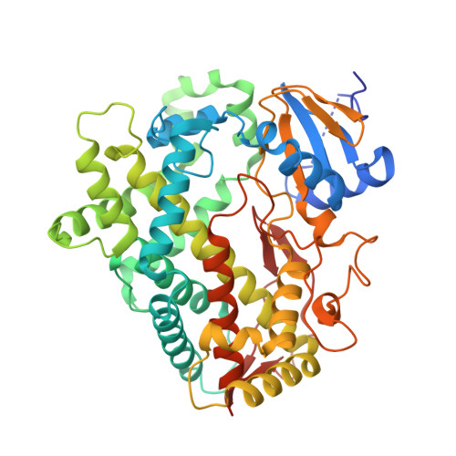
    P450 2D6 contributes significantly to the metabolism of >15% of the 200 most marketed drugs. Open and closed crystal structures of P450 2D6 thioridazine complexes were obtained using different crystallization conditions. The protonated piperidine moiety of thioridazine forms a charge-stabilized hydrogen bond with Asp-301 in the active sites of both complexes. The more open conformation exhibits a second molecule of thioridazine bound in an expanded substrate access channel antechamber with its piperidine moiety forming a charge-stabilized hydrogen bond with Glu-222. Incubation of the crystalline open thioridazine complex with alternative ligands, prinomastat, quinidine, quinine, or ajmalicine, displaced both thioridazines. Quinine and ajmalicine formed charge-stabilized hydrogen bonds with Glu-216, whereas the protonated nitrogen of quinidine is equidistant from Asp-301 and Glu-216 with protonated nitrogen H-bonded to a water molecule in the access channel. Prinomastat is not ionized. Adaptations of active site side-chain rotamers and polypeptide conformations were evident between the complexes, with the binding of ajmalicine eliciting a closure of the open structure reflecting in part the inward movement of Glu-216 to form a hydrogen bond with ajmalicine as well as sparse lattice restraints that would hinder adaptations. These results indicate that P450 2D6 exhibits sufficient elasticity within the crystal lattice to allow the passage of compounds between the active site and bulk solvent and to adopt a more closed form that adapts for binding alternative ligands with different degrees of closure. These crystals provide a means to characterize substrate and inhibitor binding to the enzyme after replacement of thioridazine with alternative compounds.


  • Organizational Affiliation

    From the Department of Molecular and Experimental Medicine and.


Macromolecules
Find similar proteins by:  (by identity cutoff)  |  3D Structure
Entity ID: 1
MoleculeChains Sequence LengthOrganismDetailsImage
Cytochrome P450 2D6
A, B
479Homo sapiensMutation(s): 0 
Gene Names: CYP2D6CYP2DL1
EC: 1.14.14.1
UniProt & NIH Common Fund Data Resources
Find proteins for P10635 (Homo sapiens)
Explore P10635 
Go to UniProtKB:  P10635
PHAROS:  P10635
GTEx:  ENSG00000100197 
Entity Groups  
Sequence Clusters30% Identity50% Identity70% Identity90% Identity95% Identity100% Identity
UniProt GroupP10635
Sequence Annotations
Expand
  • Reference Sequence
Small Molecules
Binding Affinity Annotations 
IDSourceBinding Affinity
AJN BindingDB:  4WNT Ki: min: 3.3, max: 14 (nM) from 6 assay(s)
Experimental Data & Validation

Experimental Data

  • Method: X-RAY DIFFRACTION
  • Resolution: 2.60 Å
  • R-Value Free: 0.237 
  • R-Value Work: 0.192 
  • R-Value Observed: 0.194 
  • Space Group: P 21 21 2
Unit Cell:
Length ( Å )Angle ( ˚ )
a = 57.31α = 90
b = 126.81β = 90
c = 191.73γ = 90
Software Package:
Software NamePurpose
SCALAdata scaling
PHENIXrefinement
PDB_EXTRACTdata extraction

Structure Validation

View Full Validation Report


Ligand Structure Quality Assessment 


Entry History & Funding Information

Deposition Data


Funding OrganizationLocationGrant Number
National Institutes of Health/National Institute of General Medical Sciences (NIH/NIGMS)United StatesR01GM031001

Revision History  (Full details and data files)

  • Version 1.0: 2015-01-14
    Type: Initial release
  • Version 1.1: 2015-01-21
    Changes: Database references
  • Version 1.2: 2015-03-04
    Changes: Database references
  • Version 1.3: 2017-09-13
    Changes: Author supporting evidence, Database references, Derived calculations, Other, Source and taxonomy
  • Version 1.4: 2017-11-22
    Changes: Refinement description
  • Version 1.5: 2019-12-25
    Changes: Author supporting evidence
  • Version 1.6: 2023-09-27
    Changes: Data collection, Database references, Derived calculations, Refinement description