


Crystal structure of E47Q mutant cytidine deaminase from Mycobacterium tuberculosis (MtCDA E47Q)
Sanchez-Quitian, A.Z., Rodrigues-Junior, V., Rehm, J.G., Eichler, P., Trivella, D.B.B., Bizarro, C.V., Basso, L.A., Santos, D.S.(2015) RSC Adv
Experimental Data Snapshot
wwPDB Validation 3D Report Full Report

(2015) RSC Adv
Entity ID: 1 | |||||
|---|---|---|---|---|---|
| Molecule | Chains | Sequence Length | Organism | Details | Image |
| Cytidine deaminase | 133 | Mycobacterium tuberculosis | Mutation(s): 1 EC: 3.5.4.5 | 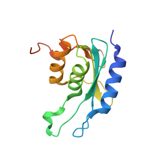 | |
UniProt | |||||
Find proteins for P9WPH3 (Mycobacterium tuberculosis (strain ATCC 25618 / H37Rv)) Explore P9WPH3 Go to UniProtKB: P9WPH3 | |||||
Entity Groups | |||||
| Sequence Clusters | 30% Identity50% Identity70% Identity90% Identity95% Identity100% Identity | ||||
| UniProt Group | P9WPH3 | ||||
Sequence AnnotationsExpand | |||||
| |||||
| Ligands 1 Unique | |||||
|---|---|---|---|---|---|
| ID | Chains | Name / Formula / InChI Key | 2D Diagram | 3D Interactions | |
| ZN Query on ZN | C [auth A], D [auth B] | ZINC ION Zn PTFCDOFLOPIGGS-UHFFFAOYSA-N |  | ||
| Length ( Å ) | Angle ( ˚ ) |
|---|---|
| a = 65.59 | α = 90 |
| b = 77.84 | β = 90 |
| c = 111.62 | γ = 90 |
| Software Name | Purpose |
|---|---|
| SCALA | data scaling |
| REFMAC | refinement |
| PDB_EXTRACT | data extraction |
RCSB PDB (citation) is hosted by
RCSB PDB is a member of the

