4WFL

Structure of the complete bacterial SRP Alu domain


Experimental Data Snapshot

  • Method: X-RAY DIFFRACTION
  • Resolution: 2.49 Å
  • R-Value Free: 0.209 
  • R-Value Work: 0.180 
  • R-Value Observed: 0.183 

wwPDB Validation   3D Report Full Report


This is version 2.0 of the entry. See complete history


Literature

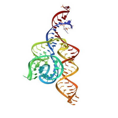
Structure of the complete bacterial SRP Alu domain.

Kempf, G.Wild, K.Sinning, I.

(2014) Nucleic Acids Res 42: 12284-12294

  • DOI: https://doi.org/10.1093/nar/gku883
  • Primary Citation of Related Structures:  
    4WFL, 4WFM

  • PubMed Abstract: 

    The Alu domain of the signal recognition particle (SRP) arrests protein biosynthesis by competition with elongation factor binding on the ribosome. The mammalian Alu domain is a protein-RNA complex, while prokaryotic Alu domains are protein-free with significant extensions of the RNA. Here we report the crystal structure of the complete Alu domain of Bacillus subtilis SRP RNA at 2.5 Å resolution. The bacterial Alu RNA reveals a compact fold, which is stabilized by prokaryote-specific extensions and interactions. In this 'closed' conformation, the 5' and 3' regions are clamped together by the additional helix 1, the connecting 3-way junction and a novel minor groove interaction, which we term the 'minor-saddle motif' (MSM). The 5' region includes an extended loop-loop pseudoknot made of five consecutive Watson-Crick base pairs. Homology modeling with the human Alu domain in context of the ribosome shows that an additional lobe in the pseudoknot approaches the large subunit, while the absence of protein results in the detachment from the small subunit. Our findings provide the structural basis for purely RNA-driven elongation arrest in prokaryotes, and give insights into the structural adaption of SRP RNA during evolution.


  • Organizational Affiliation

    Heidelberg University Biochemistry Center (BZH), INF 328, D-69120 Heidelberg, Germany.


Macromolecules
Find similar nucleic acids by:  (by identity cutoff)  |  3D Structure
Entity ID: 1
MoleculeChains LengthOrganismImage
RNA107Bacillus subtilis subsp. subtilis str. 168
Sequence Annotations
Expand
  • Reference Sequence
Small Molecules
Ligands 2 Unique
IDChains Name / Formula / InChI Key2D Diagram3D Interactions
NCO
Query on NCO

Download Ideal Coordinates CCD File 
C [auth A]
D [auth A]
F [auth A]
G [auth A]
H [auth A]
C [auth A],
D [auth A],
F [auth A],
G [auth A],
H [auth A],
I [auth A],
J [auth A],
L [auth A],
M [auth A],
N [auth A],
O [auth A],
P [auth A],
Q [auth A],
R [auth A],
S [auth A]
COBALT HEXAMMINE(III)
Co H18 N6
DYLMFCCYOUSRTK-UHFFFAOYSA-N
MG
Query on MG

Download Ideal Coordinates CCD File 
B [auth A],
E [auth A],
K [auth A]
MAGNESIUM ION
Mg
JLVVSXFLKOJNIY-UHFFFAOYSA-N
Experimental Data & Validation

Experimental Data

  • Method: X-RAY DIFFRACTION
  • Resolution: 2.49 Å
  • R-Value Free: 0.209 
  • R-Value Work: 0.180 
  • R-Value Observed: 0.183 
  • Space Group: P 21 21 21
Unit Cell:
Length ( Å )Angle ( ˚ )
a = 62.466α = 90
b = 68.283β = 90
c = 82.991γ = 90
Software Package:
Software NamePurpose
PHENIXrefinement
XDSdata reduction
Aimlessdata scaling
PHENIXphasing

Structure Validation

View Full Validation Report


Entry History & Funding Information

Deposition Data


Funding OrganizationLocationGrant Number
German Research FoundationGermanySFB638

Revision History  (Full details and data files)

  • Version 1.0: 2014-10-15
    Type: Initial release
  • Version 1.1: 2014-11-19
    Changes: Database references
  • Version 2.0: 2017-09-06
    Changes: Atomic model, Author supporting evidence, Derived calculations