4WB0

Crystal structure of the broad specificity aminotransferase from Leishmania mexicana


Experimental Data Snapshot

  • Method: X-RAY DIFFRACTION
  • Resolution: 1.91 Å
  • R-Value Free: 0.210 
  • R-Value Work: 0.177 
  • R-Value Observed: 0.178 

wwPDB Validation   3D Report Full Report


This is version 1.3 of the entry. See complete history


Literature

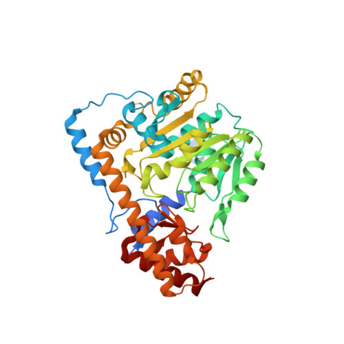
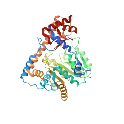
Structural basis for the relaxed substrate selectivity of Leishmania mexicana broad specificity aminotransferase.

Wen, J.Nowicki, C.Blankenfeldt, W.

(2015) Mol Biochem Parasitol 202: 34-37

  • DOI: https://doi.org/10.1016/j.molbiopara.2015.09.007
  • Primary Citation of Related Structures:  
    4WB0

  • PubMed Abstract: 

    Leishmania species are early branching eukaryotic parasites that cause difficult-to-treat tissue-damaging diseases known as leishmaniases. As a hallmark of their parasitic lifestyle, Leishmaniae express a number of aminotransferases that are involved in important cellular processes and exhibit broader substrate specificity than their mammalian host's counterparts. Here, we have determined the crystal structure of the broad specificity aminotransferase from Leishmania mexicana (LmexBSAT) at 1.91Å resolution. LmexBSAT is a homodimer and belongs to the α-branch of family-I aminotransferases. Despite the fact that the protein was crystallized in the absence of substrates and has lost the pyridoxal-5'-phosphate (PLP) cofactor during crystallization, the structure resembles the closed, ligand-bound form of related enzymes such as chicken cytosolic aspartate aminotransferase. Its broader substrate specificity seems to be rooted in increased flexibility of a substrate-binding arginine (R291) and the interactions of this residue with the N-terminus of the second chain of the dimer.


  • Organizational Affiliation

    Technische Universität Dortmund, Fakultät Chemie, Otto-Hahn-Str. 6, 44227 Dortmund, Germany; Physical Biochemistry, Max Planck Institute for Molecular Physiology, Otto-Hahn-Str. 11, 44227 Dortmund, Germany.


Macromolecules
Find similar proteins by:  (by identity cutoff)  |  3D Structure
Entity ID: 1
MoleculeChains Sequence LengthOrganismDetailsImage
Broad specificity aminotransferase414Leishmania mexicanaMutation(s): 0 
UniProt
Find proteins for Q71PB4 (Leishmania mexicana)
Explore Q71PB4 
Go to UniProtKB:  Q71PB4
Entity Groups  
Sequence Clusters30% Identity50% Identity70% Identity90% Identity95% Identity100% Identity
UniProt GroupQ71PB4
Sequence Annotations
Expand
  • Reference Sequence
Find similar proteins by:  (by identity cutoff)  |  3D Structure
Entity ID: 2
MoleculeChains Sequence LengthOrganismDetailsImage
Broad specificity aminotransferase414Leishmania mexicanaMutation(s): 0 
UniProt
Find proteins for Q71PB4 (Leishmania mexicana)
Explore Q71PB4 
Go to UniProtKB:  Q71PB4
Entity Groups  
Sequence Clusters30% Identity50% Identity70% Identity90% Identity95% Identity100% Identity
UniProt GroupQ71PB4
Sequence Annotations
Expand
  • Reference Sequence
Small Molecules
Modified Residues  1 Unique
IDChains TypeFormula2D DiagramParent
CSO
Query on CSO
A
L-PEPTIDE LINKINGC3 H7 N O3 SCYS
Experimental Data & Validation

Experimental Data

  • Method: X-RAY DIFFRACTION
  • Resolution: 1.91 Å
  • R-Value Free: 0.210 
  • R-Value Work: 0.177 
  • R-Value Observed: 0.178 
  • Space Group: P 21 21 21
Unit Cell:
Length ( Å )Angle ( ˚ )
a = 86.418α = 90
b = 90.564β = 90
c = 142.159γ = 90
Software Package:
Software NamePurpose
PHENIXrefinement
XDSdata reduction
Aimlessdata scaling
PHASERphasing

Structure Validation

View Full Validation Report


Entry History 

Deposition Data

Revision History  (Full details and data files)

  • Version 1.0: 2015-09-09
    Type: Initial release
  • Version 1.1: 2015-11-25
    Changes: Database references
  • Version 1.2: 2018-04-25
    Changes: Data collection
  • Version 1.3: 2024-01-10
    Changes: Data collection, Database references, Refinement description