


Two synonymous gene variants encode proteins with identical sequence, but different folding conformations.
Buhr, F., Jha, S., Thommen, M., Rodnina, M., Kutz, F., Kudlinzki, D., Linhard, V.L., Komar, A.A., Schwalbe, H.To be published.
Experimental Data Snapshot
wwPDB Validation 3D Report Full Report

Entity ID: 1 | |||||
|---|---|---|---|---|---|
| Molecule | Chains | Sequence Length | Organism | Details | Image |
| Gamma-crystallin B | 181 | Bos taurus | Mutation(s): 0 Gene Names: CRYGB | 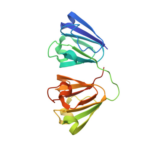 | |
UniProt | |||||
Find proteins for P02526 (Bos taurus) Explore P02526 Go to UniProtKB: P02526 | |||||
Entity Groups | |||||
| Sequence Clusters | 30% Identity50% Identity70% Identity90% Identity95% Identity100% Identity | ||||
| UniProt Group | P02526 | ||||
Sequence AnnotationsExpand | |||||
| |||||
| Length ( Å ) | Angle ( ˚ ) |
|---|---|
| a = 123.641 | α = 90 |
| b = 123.641 | β = 90 |
| c = 56.548 | γ = 120 |
| Software Name | Purpose |
|---|---|
| PHENIX | refinement |
| XDS | data reduction |
| Coot | model building |
| PHASER | phasing |
| XSCALE | data scaling |
RCSB PDB (citation) is hosted by
RCSB PDB is a member of the

