4W97

Structure of ketosteroid transcriptional regulator KstR2 of Mycobacterium tuberculosis


Experimental Data Snapshot

  • Method: X-RAY DIFFRACTION
  • Resolution: 1.60 Å
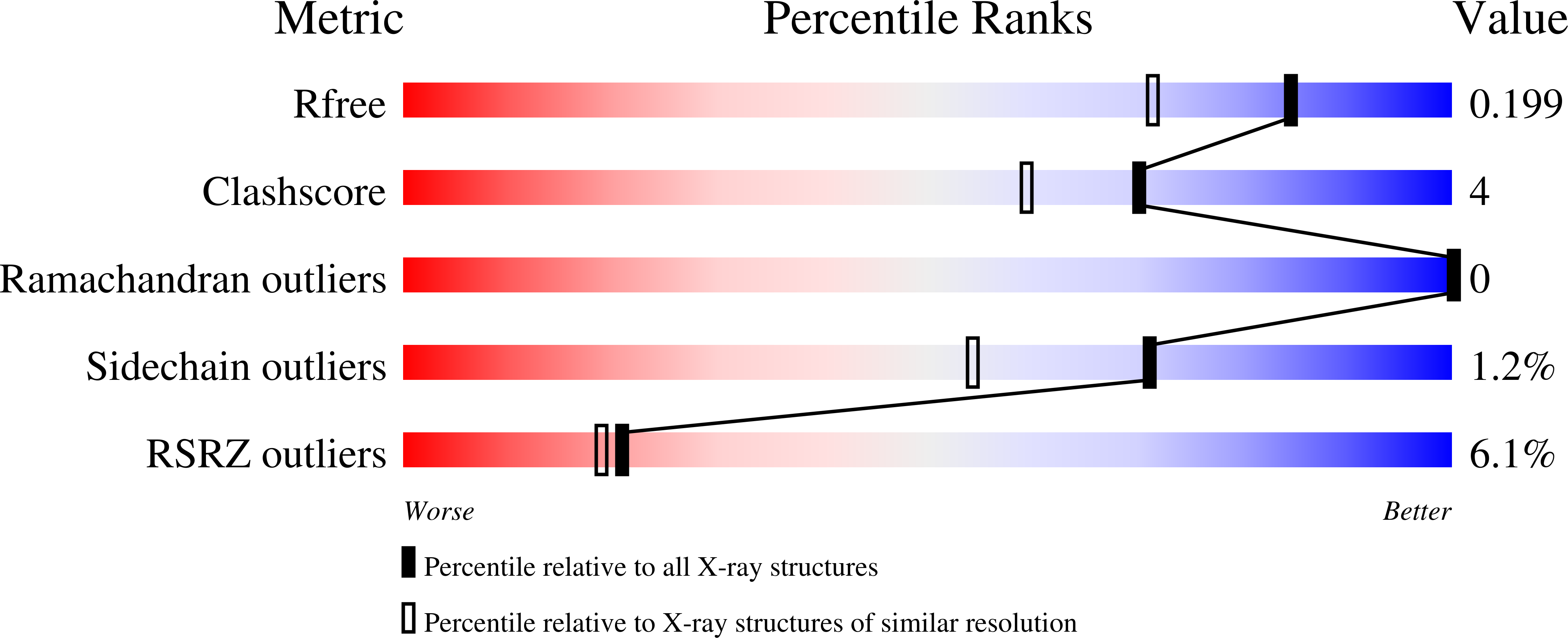
  • R-Value Free: 0.199 
  • R-Value Work: 0.160 
  • R-Value Observed: 0.162 

wwPDB Validation   3D Report Full Report


Ligand Structure Quality Assessment 


This is version 2.0 of the entry. See complete history


Literature

Structural and Functional Characterization of a Ketosteroid Transcriptional Regulator of Mycobacterium tuberculosis.

Crowe, A.M.Stogios, P.J.Casabon, I.Evdokimova, E.Savchenko, A.Eltis, L.D.

(2015) J Biol Chem 290: 872-882

  • DOI: https://doi.org/10.1074/jbc.M114.607481
  • Primary Citation of Related Structures:  
    4W97

  • PubMed Abstract: 

    Catabolism of host cholesterol is critical to the virulence of Mycobacterium tuberculosis and is a potential target for novel therapeutics. KstR2, a TetR family repressor (TFR), regulates the expression of 15 genes encoding enzymes that catabolize the last half of the cholesterol molecule, represented by 3aα-H-4α(3'-propanoate)-7aβ-methylhexahydro-1,5-indane-dione (HIP). Binding of KstR2 to its operator sequences is relieved upon binding of HIP-CoA. A 1.6-Å resolution crystal structure of the KstR2(Mtb)·HIP-CoA complex reveals that the KstR2(Mtb) dimer accommodates two molecules of HIP-CoA. Each ligand binds in an elongated cleft spanning the dimerization interface such that the HIP and CoA moieties interact with different KstR2(Mtb) protomers. In isothermal titration calorimetry studies, the dimer bound 2 eq of HIP-CoA with high affinity (K(d) = 80 ± 10 nm) but bound neither HIP nor CoASH. Substitution of Arg-162 or Trp-166, residues that interact, respectively, with the diphosphate and HIP moieties of HIP-CoA, dramatically decreased the affinity of KstR2(Mtb) for HIP-CoA but not for its operator sequence. The variant of R162M that decreased the affinity for HIP-CoA (ΔΔG = 13 kJ mol(-1)) is consistent with the loss of three hydrogen bonds as indicated in the structural data. A 24-bp operator sequence bound two dimers of KstR2. Structural comparisons with a ligand-free rhodococcal homologue and a DNA-bound homologue suggest that HIP-CoA induces conformational changes of the DNA-binding domains of the dimer that preclude their proper positioning in the major groove of DNA. The results provide insight into KstR2-mediated regulation of expression of steroid catabolic genes and the determinants of ligand binding in TFRs.


  • Organizational Affiliation

    From the Departments of Biochemistry and Molecular Biology and.


Macromolecules
Find similar proteins by:  (by identity cutoff)  |  3D Structure
Entity ID: 1
MoleculeChains Sequence LengthOrganismDetailsImage
HTH-type transcriptional repressor KstR2200Mycobacterium tuberculosis H37RvMutation(s): 0 
Gene Names: kstR2Rv3557c
UniProt
Find proteins for P9WMB9 (Mycobacterium tuberculosis (strain ATCC 25618 / H37Rv))
Explore P9WMB9 
Go to UniProtKB:  P9WMB9
Entity Groups  
Sequence Clusters30% Identity50% Identity70% Identity90% Identity95% Identity100% Identity
UniProt GroupP9WMB9
Sequence Annotations
Expand
  • Reference Sequence
Small Molecules
Ligands 2 Unique
IDChains Name / Formula / InChI Key2D Diagram3D Interactions
UCA
Query on UCA

Download Ideal Coordinates CCD File 
B [auth A]S-[2-[3-[[(2R)-4-[[[(2R,3S,4R,5R)-5-(6-aminopurin-9-yl)-4-oxidanyl-3-phosphonooxy-oxolan-2-yl]methoxy-oxidanyl-phosphoryl]oxy-oxidanyl-phosphoryl]oxy-3,3-dimethyl-2-oxidanyl-butanoyl]amino]propanoylamino]ethyl] 3-[(3aS,4S,7aS)-7a-methyl-1,5-bis(oxidanylidene)-2,3,3a,4,6,7-hexahydroinden-4-yl]propanethioate
C34 H52 N7 O19 P3 S
IWNWMTZIJPUDPV-MDQHZGBLSA-N
CL
Query on CL

Download Ideal Coordinates CCD File 
C [auth A],
D [auth A],
E [auth A],
F [auth A]
CHLORIDE ION
Cl
VEXZGXHMUGYJMC-UHFFFAOYSA-M
Binding Affinity Annotations 
IDSourceBinding Affinity
UCA Binding MOAD:  4W97 Kd: 80 (nM) from 1 assay(s)
Experimental Data & Validation

Experimental Data

Unit Cell:
Length ( Å )Angle ( ˚ )
a = 72.485α = 90
b = 90.476β = 129.68
c = 49.798γ = 90
Software Package:
Software NamePurpose
PHENIXrefinement
StructureStudiodata collection
HKL-3000data reduction
HKL-3000data scaling
MrBUMPphasing
PHENIXmodel building

Structure Validation

View Full Validation Report


Ligand Structure Quality Assessment 


Entry History 

Deposition Data

Revision History  (Full details and data files)

  • Version 1.0: 2014-11-26
    Type: Initial release
  • Version 1.1: 2014-12-03
    Changes: Database references
  • Version 1.2: 2015-01-21
    Changes: Database references
  • Version 2.0: 2023-09-27
    Changes: Atomic model, Data collection, Database references, Derived calculations, Other, Refinement description, Source and taxonomy