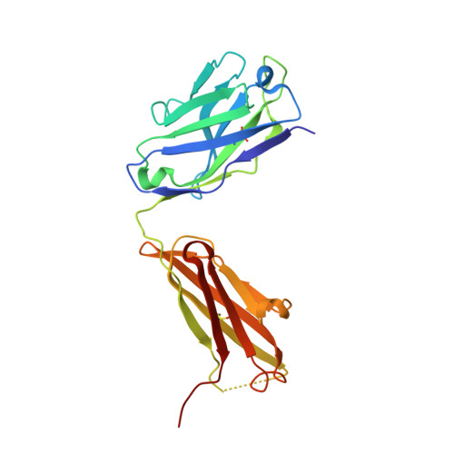
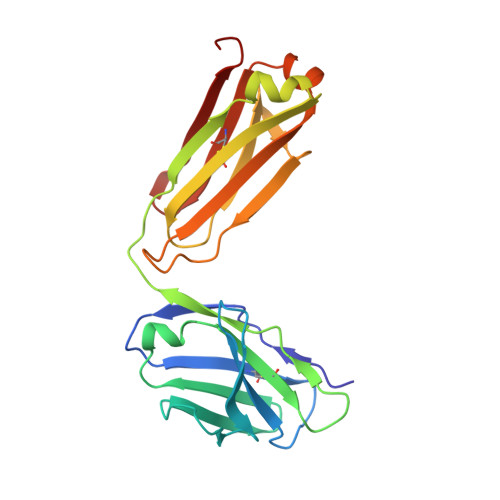
Structural Basis for Quinine-Dependent Antibody Binding to Platelet Integrin Alphaiib Beta3
Zhu, J., Zhu, J., Bougie, D.W., Aster, R.H., Springer, T.A.(2015) Blood 126: 2138
- PubMed: 26282540
- DOI: https://doi.org/10.1182/blood-2015-04-639351
- Primary Citation of Related Structures:
4UIK, 4UIL, 4UIM, 4UIN - PubMed Abstract:
Drug-induced immune thrombocytopenia (DITP) is caused by antibodies that react with specific platelet-membrane glycoproteins when the provoking drug is present. More than 100 drugs have been implicated as triggers for this condition, quinine being one of the most common. The cause of DITP in most cases appears to be a drug-induced antibody that binds to a platelet membrane glycoprotein only when the drug is present. How a soluble drug promotes binding of an otherwise nonreactive immunoglobulin to its target, leading to platelet destruction, is uncertain, in part because of the difficulties of working with polyclonal human antibodies usually available only in small quantities. Recently, quinine-dependent murine monoclonal antibodies were developed that recognize a defined epitope on the β-propeller domain of the platelet integrin αIIb subunit (GPIIb) only when the drug is present and closely mimic the behavior of antibodies found in human patients with quinine-induced thrombocytopenia in vitro and in vivo. Here, we demonstrate specific, high-affinity binding of quinine to the complementarity-determining regions (CDRs) of these antibodies and define in crystal structures the changes induced in the CDR by this interaction. Because no detectable binding of quinine to the target integrin could be demonstrated in previous studies, the findings indicate that a hybrid paratope consisting of quinine and reconfigured antibody CDR plays a critical role in recognition of its target epitope by an antibody and suggest that, in this type of drug-induced immunologic injury, the primary reaction involves binding of the drug to antibody CDRs, causing it to acquire specificity for a site on a platelet integrin.
Organizational Affiliation:
Department of Biological Chemistry and Molecular Pharmacology, Harvard Medical School, Boston, MA; Program in Cellular and Molecular Medicine, Children's Hospital Boston, Boston, MA;